What are key parts of the social brain circuit used in social processing?
Researchers have found a positive correlation between circulating dendritic cell number and amygdala size—the more circulating dendritic cells, the bigger the amygdala. To learn more about the amygdala and other brain structures involved in the social brain circuit, please read below.
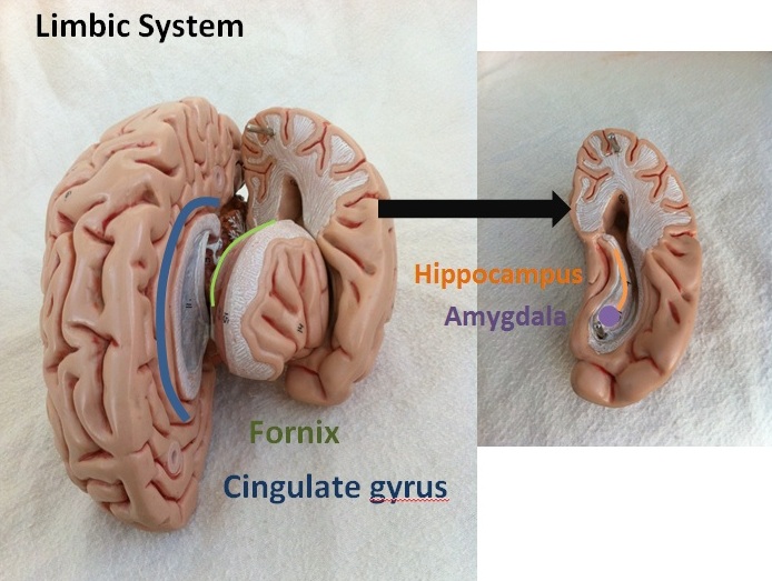
Scientists have identified the social circuit in the brain by means of brain imaging, electrical stimulation, and lesions in other animals. Imaging the brains of people watching animated movies of simple geometric shapes interacting in a “social,” mechanical,” or “random” way has identified the amygdala, fusiform gyrus, prefrontal cortex, and superior temporal sulcus as some of the key parts of the social pathway.1,2 Many key regions belong to the limbic system, which regulates emotions. The limbic system is also connected to our autonomic nervous system, which regulates our internal organs and affects how our body deals with stress.
 The amygdala attaches emotional significance to sensory stimuli, along with being an important site for learning and memory. Some cells in the amygdala (and other brain regions) contain key hormones, or neuropeptides, that play a role in social behavior: oxytocin and vasopressin. These neuropeptides are important in social recognition, social contact, and social bonding; if these peptides are injected into the nose, they can reduce social stress and enhance positive social feelings such as trust.3 Males are more dependent than females on vasopressin for social behavior, and the hormone testosterone can change vasopressin’s activity. Interestingly, males are also four times as likely as females to develop ASD. Other important brain regions linking social and environmental cues with hormonal control (including the neuropeptides vasopressin and oxytocin, along with testosterone and estrogen) are the pre-optic area and hypothalamus.
The fusiform gyrus, located underneath the amygdala, helps us recognize and process faces. The prefrontal cortex, part of our frontal cortex, is connected to the amygdala and is involved in problem solving, emotion, and complex thought.
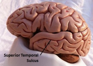
 The superior temporal sulcus contains one of nature’s most interesting neuronal classes: mirror neurons. In humans, these neurons “mirror” or fire when other people perform an action as well as show their emotions—anything from kicking a soccer ball to crying. These same neurons are activated when we perform the same action or experience the same emotion ourselves.4
When we talk, smile, or physically interact with others, their positive response to our behavior encourages us to remain engaged, or socially motivated. We gain pleasure from the interaction through our dopamine reward pathway.5 When we are socially engaged, such as when making eye contact, the neurotransmitter dopamine is released.
| References: |
|

