

How brains are imaged
This study looked at the activity of brains in ASD patients while being provided with either social or monetary rewards. The specific method used in this study to measure the activity of particular brain areas was fMRI. Read more below about two other methods that could have been used to measure activity.

What can be measured in a brain?
Researchers measure four different components in human brain studies:
- The volumes and positions of the different brain areas.
- The interconnections between different brain areas.
- The activity of a particular brain area.
- The chemical composition of a brain area.
1. Measuring volumes and positions of different brain areas
If you were asked to find differences in the brain of a person diagnosed with ASD, probably your first approach would be to look at brains and see whether there are any differences between an individual with ASD and a neurotypical person. Although this can be done after a person dies, it is more common to do this while the person is still alive. The workhorse for this approach has been a technique called magnetic resonance imaging (MRI).1
2. Measuring interconnections between different brain areas
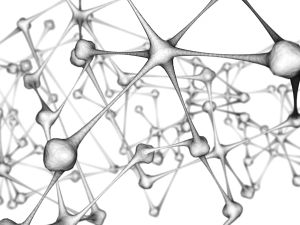
In the early 1990s, some researchers found a way to slightly modify the MRI technique to measure connections between areas.2 This modified technique is called diffusion tensor imaging (DTI). The number of studies using this technique is growing rapidly, and we are now at the beginning of an era of connectivity studies in the human brain.
3. Measuring the activity of a particular brain area
Neurons use a lot of energy and oxygen when they are busy processing information. The brain appears to be compartmentalized in some way so that different areas process different types of information. So, at least in theory, if you can measure where energy is being used while a person is performing a certain task, then you can deduce what each brain area does. And for people with ASD, perhaps you could find areas that are not being used when they should be.
4. Measuring the chemical composition of a brain area
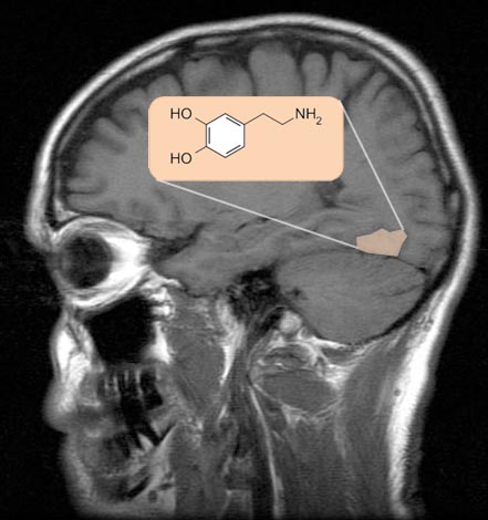
Neurons function by communicating with other neurons. One of the ways that a neuron can "talk" to another neuron is by transferring a specific chemical, or neurotransmitter. When researchers looked at different brain areas, they found that each area had a specific set of neurotransmitters that was different from those of other brain areas. The set of neurotransmitters found in a brain area affects the types of messages it can send to the rest of the brain.
| References: |
|