

Categories
Select a new category
Brain Study Reveals Commonality between ASD and ADHD
By Chelsea Toledo, M.A. on July 29, 2018

Background: Autism Spectrum Disorder (ASD) is a neurodevelopmental condition characterized by differences in communication, behavior, and social development. Prior brain-imaging studies have shown pattered differences in the connectivity of brains of people with ASD compared to their typically developing peers. However, little research has compared the brains of ASD with those of people with other neurodevelopmental disorders.
What’s New: A new study compared the connectivity of brains of people with ASD to those of people with attention deficit/hyperactivity disorder (ADHD), seeking commonalities. The researchers leveraged existing brain-imaging data from a total of 1305 people between the ages of 7 and 21 – 284 with ASD, 369 with ADHD, and 652 age-matched controls with typical development.
The researchers found:
- Three main factors were common to the brains of people with ASD and ADHD, but not people with typical development.
- The three factors were differences in the connectivity of the default mode network (a set of brain regions active when a person is daydreaming), the dorsal attention network (a set of brain regions active when a person is looking into space), and the salience network (a set of brain regions active in identifying important stimuli).
- These patterns likely comprise a “neural signature” for people with ASD and those with ADHD, reinforcing existing diagnoses made for either disorder.
Why it’s important: This study reveals underlying patterns in brain connectivity in both people with ASD and those with ADHD. Further research could help define this “neural signature” for clinical application.
Image Credit: Nevit Dilmen, NIH 3D Print Exchange, National Institutes of Health. Shared via Flickr under a Creative Commons license.
Help me understand :
| Source(s) : |
| Tweet |
Inexpensive Brain Scans Could Predict Autism Spectrum Disorder
By Chelsea Toledo, M.A. on May 23, 2018
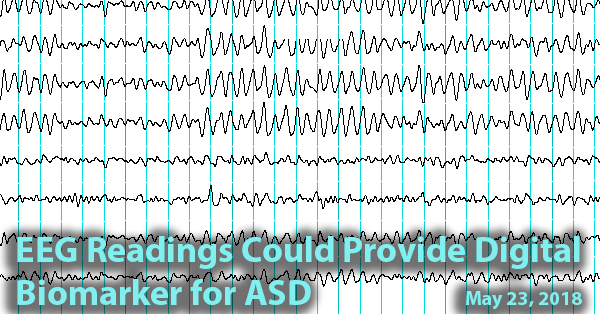
Background: A reliable biomarker for early detection of autism is critical. Electroencephalography (EEG), a technique for recording and mapping electrical activity in the brain – could provide such a measure. During this non-invasive and relatively inexpensive procedure, small metal discs called electrodes are placed on the scalp to detect fluctuations in the voltage given off by the brain’s neurons over time.
What’s New: In a new study, researchers assessed whether EEG data could accurately predict emergence of Autism Spectrum Disorder (ASD) in very young children. They performed EEG procedure on 99 high-risk children (defined as having an older sibling with ASD) and 89 age-matched controls up to seven times, beginning when the children were as young as 3 months old and ending when the children were 3 years old.
The researchers found:
- Patterns in EEG data allowed researchers to predict nearly 100 percent of ASD cases prospectively diagnosed using the Autism Diagnostic Observation Schedule (ADOS) at either 18, 24, or 36 months of age.
- The EEG data also helped predict the severity of ASD as early as 3 months of age.
- Significant differences were evident between the EEG data of the high-risk versus the low-risk group. These differences were most pronounced at 12 months of age.
Why it’s important: This study suggests that EEG could be a useful tool, along with behavioral analyses, for diagnosing ASD early. Research has shown that early diagnosis is associated with improved outcomes for children with ASD.
Help me understand :
| Source(s) : |
| Tweet |
Neurofeedback Training Improves Brain Connectivity in People with ASD
By Chelsea Toledo, M.A. on April 17, 2018

Background: In recent years, researchers and clinicians have begun to leverage on brain imaging technology to provide “neurofeedback.” Through this process, they image the brain for aberrant activity and provide positive feedback when desired activity in that area is achieved. That positive feedback can take the form of a green light or another positive signal to participants.
What’s New: A recent study assessed the effectiveness of neurofeedback using functional magnetic resonance imaging, or fMRI, in young adult and adolescent males with ASD. The researchers conducted four training sessions over a period of eight days in 17 males with ASD, between the ages of 15 and 25, alongside 11 age-matched controls with typical development. Each training consisted of a series of “rest scans” with no task for the participant, along with a series of “puzzle tasks,” during which the participants attempted to reveal a hidden picture underneath a blank screen. When the participants exhibited the desired brain activity, positive feedback was given via revealing part of the picture and playing an upbeat sound.
The researchers found:
- The training resulted in improved connectivity in the whole brain, with the greatest differences observed in connectivity between two regions of interest, which previous research has linked to ASD. These changes were not found in the control group.
- The observed improvements in connectivity continued in subsequent “rest scans.”
- The improvements in brain connectivity correlated with improvements in the ASD participants’ behavior, as assessed by behavioral questionnaires filled out by parents before and after the series of trainings.
Why it’s important: This study provides further evidence of the role of brain connectivity in ASD. It also points to a potentially effective and non-invasive treatment option to address related behavioral issues. Larger studies including other participants (e.g., children and female groups) could further explore this possibility.
Help me understand :
| Source(s) : |
| Tweet |
Extra Stable Brain Activity Patterns Found in ASD
By Shana R. Spindler, PhD and Chelsea E. Toledo, M.A. on August 14, 2017
Background: Even at rest, the brain never really rests. Brain cells, or neurons, use chemicals to shoot information at lightening speed throughout the brain all the time. This brain activity occurs in special patterns that are related to the different functions of the brain. Scientists think that measuring patterns of brain activity during a resting state can offer clues about why some people have autism.
What’s new: On July 5, 2017, the journal Nature Communications released a study comparing resting brain activity in typically developing individuals versus their peers with high-functioning autism spectrum disorder (ASD). Researchers looked at brain imaging data taken in a resting state from a total of 50 male individuals between the ages of 18 and 39.
The researchers used a special type of mathematical formula to make brain activity look like peaks and valleys on a topographical map. They found that individuals with ASD had differently sized peaks and valleys as compared to their typically developing peers. This particular method of analyzing brain activity allowed autism identification—based solely on brain scans—with about 85% accuracy. The researchers also found a correlation between the peak and valley sizes and the severity of autism symptoms.
Why it’s important: The study is an important step forward in establishing ways to extract information from brain scans, which in this case was a functional magnetic resonance imaging (fMRI) scan. The findings of the study suggest a stability of brain activity pattern that differs between typically developing individuals and those with autism spectrum disorder. These differences may one day aid autism diagnosis or indicate severity along the spectrum, but additional investigation is required.
Help me understand :
| Source(s) : |
| Tweet |
New Brain-Imaging Study Identifies Autism in Infants
By Chelsea E. Toledo, M.A. on March 7, 2017
Background: Early diagnosis of Autism Spectrum Disorder (ASD) is a topic of great interest to parents and researchers alike. Studies have repeatedly shown that the earlier a child received intervention for ASD, the more benefits that child will gain related to the behavioral, communicative, and social symptoms of the disorder. However, in the United States – where one in 68 children receives an ASD diagnosis – the average age of diagnosis is 4 years old. Furthermore, standard practices aren’t yet in place for clinicians to diagnose ASD before 2 years old, when a great deal of the development of language and social skills has already taken place.
What’s new: On February 16, 2017, the journal Nature published a study outlining a potential measure for predicting ASD diagnosis before symptoms are apparent in young children. The researchers performed brain imaging at three different points in time on a total of 148 infants – 42 of whom defined as low-risk and 106 of whom were defined as high-risk, based on whether or not the infants had older siblings with ASD. Measuring the growth of the brain’s surface area at 6, 12, and 24 months, the researchers noted a trend in accelerated growth (especially in the brain’s cortex, which processes information from the environment) in the infants who were ultimately diagnosed with ASD. The researchers used this information to develop an algorithm that accurately predicted 80 percent of ASD cases in a separate group of infants.
Why it’s important: This is the first study to follow the same children from infancy to toddlerhood, tracing risk factors for diagnosis to actual clinical outcomes. Future studies could combine the researchers’ algorithm with other useful predictors (such as genetics and behavior) to improve its accuracy, with the aim of developing an early diagnostic technique.
Help me understand :
| Source(s) : |
| Tweet |
Motor Deficits with ASD Linked to Right Side of Brain
By Shana R. Spindler, PhD on August 10, 2016
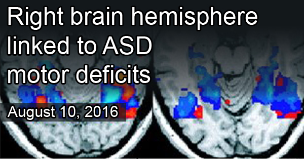
Background: At least 80% of children with autism spectrum disorder (ASD) have motor problems, according to recent estimates. Typical problems include delayed motor skills and trouble with coordination, such as kicking a ball or grasping small objects. In some cases, motor problems are apparent before other ASD symptoms.
What’s new: A new brain imaging study looked at motor control in the left and right sides of the brain in 8 to12 year old children with ASD. Researchers visualized brain activity, during a finger-tapping task, of 44 children with high-functioning autism and 80 typical, control children. Normally, regions in the left side of the brain specialize in language and motor functions. Previous work found that these regions are right-side dominant for language processing in children with ASD. Similarly, in this study researchers found brain regions that were right-side dominant for motor control too.
Why it’s important: This is the first study to look at left- and right-brain activity related to motor functions in children with ASD. The finding that some motor control is shifted to the right side of the brain in ASD is important given the potential for brain imaging to provide markers for early diagnosis.
This work was published on July 14, 2016, in the journal Molecular Autism.
Help me understand :
| Source(s) : |
| Tweet |
Language Delay in ASD Linked to Brain Symmetry
By Chelsea E. Toledo, M.A. on March 29, 2016
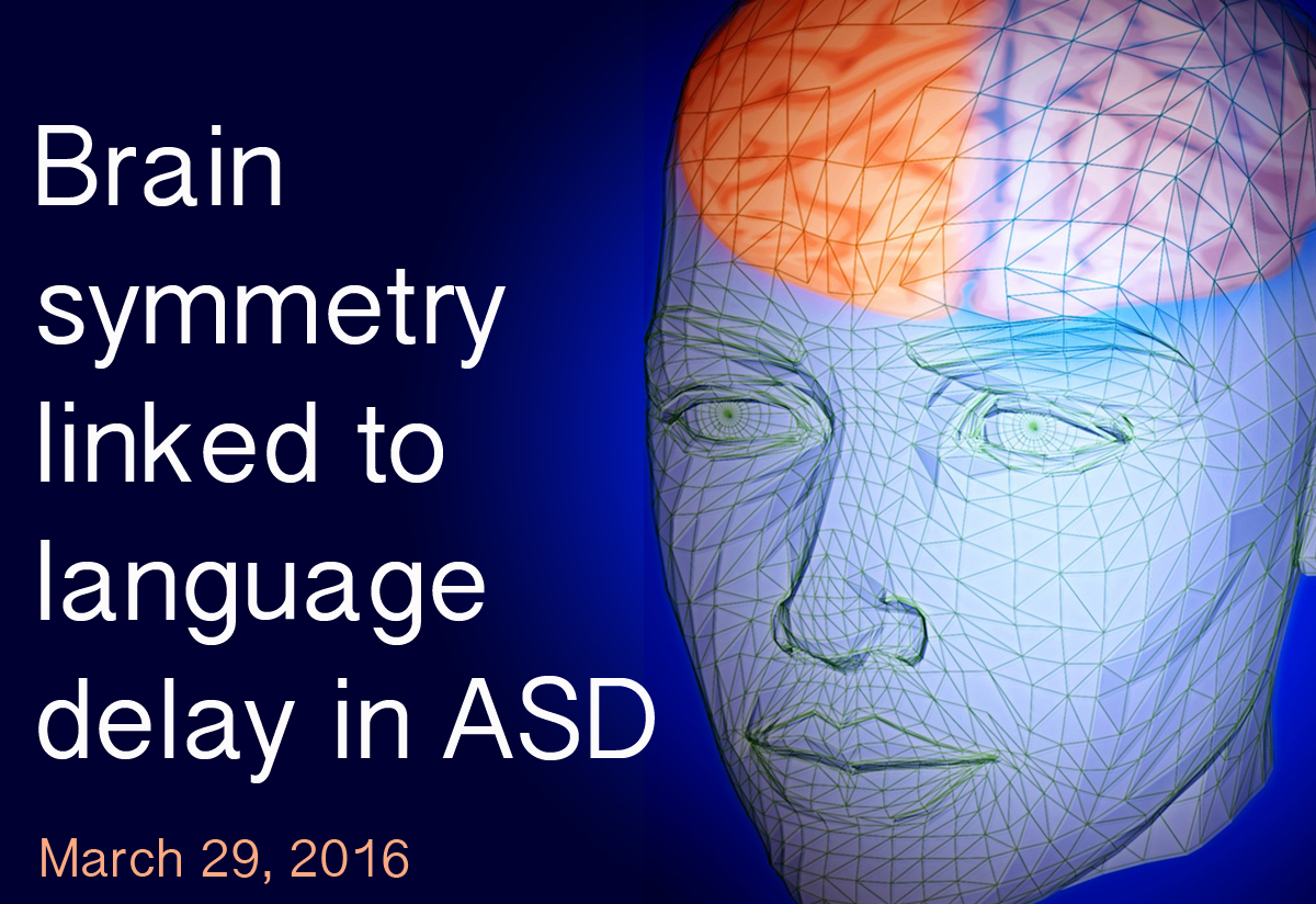
Background: Autism spectrum disorder (ASD) encompasses a broad array of conditions characterized by differences in socialization, communication, and behavior. One area where people with ASD can differ widely from one another is in language acquisition. Some individuals face severe delays in speech while others progress more typically.
What’s New: The January 2016 issue of Human Brain Mapping featured a study that explored the symmetry between brain hemispheres in individuals with or without language delay – defined as uttering one’s first words after 24 months of age or first phrases after 33 months of age. The researchers performed structural magnetic resonance imaging (MRI) on 136 right-handed men between the ages of 18 and 43 (67 with ASD and 69 with typical development). Participants in the ASD group had reduced asymmetry between several brain areas across the left and right sides of the brain, which was most pronounced in those with language delay and/or abnormal social functioning.
Why it’s important: This structural asymmetry of the brain could eventually help clinicians diagnose and understand specific subgroups of ASD. Future research could examine the brain symmetry of women and/or left-handed individuals with ASD.
Help me understand :
| Source(s) : |
| Tweet |
Study Probes Normalized Symptoms of Autism
By Chelsea E. Toledo, M.A. on March 22, 2016
Background: Autism spectrum disorder (ASD) is generally characterized by differences in socialization, communication, and behavior. Research has shown differences in brain activity underlying these traits. However, certain individuals have been shown to “grow out” of ASD symptoms, with social, communicative, and behavioral traits mirroring those of their typically developing peers. Little is known about this phenomenon—do individuals experience suppression of atypical brain activity, or does the brain establish compensatory mechanisms?
What’s New: On December 2, 2015, the journal NeuroImage: Clinical published a study exploring the underlying brain activity of people with ASD whose symptoms became normalized later in life. The researchers administered a sentence comprehension task while performing brain scans on 59 participants between the ages of 8 and 21 (23 with high-functioning ASD, 20 with typical development, and 16 who no longer met the criteria for an ASD diagnosis). They found similar activity between the ASD and normalized ASD groups, with enhanced compensatory brain activation in the group with normalized symptoms.
Why it’s important: This study suggests that people with ASD do not “grow out” of the disorder, as their brain activity more closely mirrors that of people with ASD than those with typical development when processing language. Instead, the brain may compensate by increasing activity in language processing areas of the brain. Future studies could investigate patterns of brain activity when completing other types of tasks, and additional research may reveal the role of different types of therapy in ameliorating ASD symptoms.
Help me understand :
| Source(s) : |
| Tweet |
Brain Imaging Far from ASD Diagnosis, But Getting There
By Shana R. Spindler, Ph.D. on February 16, 2016
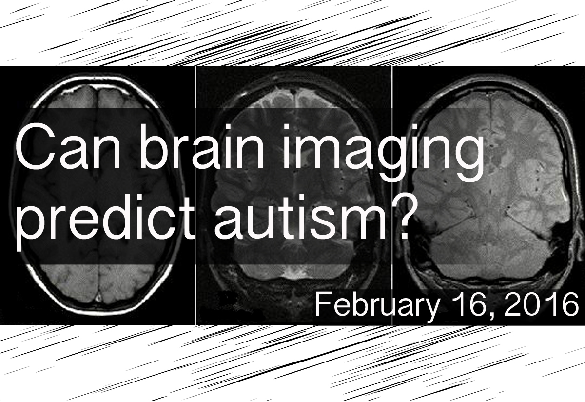
Background: A major goal in autism research is to identify measurable indicators, known as biomarkers, for Autism Spectrum Disorder (ASD) diagnosis. To date, researchers have struggled to find a universal commonality among the brains of individuals with autism. Magnetic Resonance Imaging (MRI), a type of scan that uses magnetic fields and radio waves to create detailed images of organ systems, is a popular technique to study brain structures in a living person and holds much promise for finding an ASD biomarker.
What’s new: At the 37th International Conference of the Institute of Electrical and Electronics Engineers, researchers reported on how well structural MRI predicts an autism diagnosis. In a large study using a well-established set of MRI brain scans from 15 research centers across the United States, researchers performed several experiments, showing that:
- The accuracy of autism classification by MRI got better with increased autism severity
- MRI detected possible anatomical features of autism with higher sensitivity when the individual was less than ten years of age or more than 30 years of age
- The frontal and temporal regions of the brain were important for autism classification by MRI
- Due to the heterogeneous nature of ASD, studies using a small number of participants may inflate the actual predictive power of MRI
Why it’s important: This is the first study to find that autism severity, according to social and communicative behaviors, correlates with the predictive power of MRI. The results suggest that individual behavioral information may help augment MRI findings. The study also highlights the importance of social and language regions of the brain, key age ranges for MRI effectiveness, and the requirement for large numbers of participants in brain imaging studies for autism.
Help me understand :
| Source(s) : |
| Tweet |
Regressive ASD Linked to Temporal Lobe Activity
By Chelsea E. Toledo, M.A. on September 1, 2015
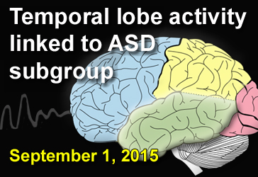
Background: The temporal lobe, one of the four major parts of the brain’s cerebral cortex, aids in processing sound, categorizing objects, and consolidating memories. It also plays a role in visual processing, emotions, and language. Researchers first began to suspect a relationship between temporal lobe abnormalities and autism spectrum disorder (ASD) in 1975. (see second Pubmed link below)
What’s New: On July 30, 2015, the journal European Child & Adolescent Psychiatry published a study investigating if irregular electrical brain activity – an indicator of epilepsy – could predict any specific feature of ASD. The researchers analyzed the results of brain scans from 220 children and young adults, 71 with ASD and 146 with other developmental disorders. After performing brain measurements on a subgroup of participants, they found a correlation between abnormal electrical brain activity, relatively large head size, and loss of previously acquired developmental skills, known as regression. The relationship between regression and abnormal electrical brain activity was the strongest when that activity took place in the temporal lobe.
Why it’s important: The results of this study support the possibility that irregular electrical activity in distinct brain areas may predict or help define ASD subgroups.
Help me understand :
| Source(s) : |
| Tweet |
Study Pinpoints Differences in Autistic Brain
By Chelsea E. Toledo, M.A. on March 31, 2015
Background: Autism Spectrum Disorder (ASD) is characterized by differences in social interaction, communication, and behavior. Researchers have hypothesized that the functioning of—and communication between—related brain regions is atypical in people affected by the disorder. Their studies have largely focused on select areas of the brain, as opposed to observing them within the context of the entire brain system.
What’s new: On March 20, 2015, the journal Brain published a large study providing a whole-brain perspective of ASD versus typical development. The researchers used a grid-based technique to analyze the entire brain at once with data from existing functional Magnetic Resonance Imaging (fMRI) scans of the resting brain. They found that, in comparison to 509 typically developing individuals, the 419 participants with ASD showed differences in 20 key areas.
Why it’s important: The observed differences were in areas of the brain active in processing facial expressions, sense of self, and theory of mind—the ability to discern and predict other’s mental states. Future studies could leverage this technique to learn more about brain function in obsessive compulsive disorder, attention deficit hyperactive disorder, and schizophrenia.
Help me understand :
| Source(s) : |
| Tweet |
Empathy For Others in Pain Present in ASD
By Shana R. Spindler, Ph.D. on January 23, 2014
Background: Empathy is the ability to understand what another person is feeling. The lack of this trait was thought to underlie some of the social problems present in people with Autism Spectrum Disorder (ASD). However, few studies have addressed the brain activity of those with ASD in situations where empathy would be likely.
What’s new: On January 14, 2014, the online journal Translational Psychiatry published a study that suggests people with ASD maintain the neural ability to process empathy, at least when seeing others in pain. Researchers used functional magnetic resonance imaging to examine the spontaneous brain activity of 38 adolescents and adults with high-functioning ASD and 35 age-matched controls as they watched two-second videos of people with shoulder pain.
Contrary to popular belief, the individuals with ASD showed no significant difference in spontaneous brain activity upon seeing someone in pain as compared to the control group. Both groups had activity in brain areas involving emotional arousal and understanding. At a lower statistical threshold, the group with ASD appeared to have increased activity in brain areas likely used for situation assessment.
Why it’s important: The researchers suggest that empathy remains intact in ASD, while the perceived lack of caring links to attempts to calm personal distress by reassessing the situation. The authors of the study note that pain is a very basic social cue and that future studies should look at additional situations where empathy is used.
Help me understand :
| Source(s) : |
| Tweet |
Oxytocin Alters Amygdala Activity to Social Stimuli
By Shana R. Spindler, PhD on April 11, 2013
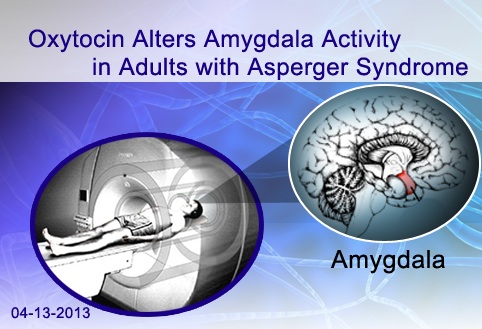
Background: Oxytocin is a hormone that is produced in the brain and released into the bloodstream and central nervous system, where it binds to oxytocin receptors on cells. Its role in helping labor contractions was discovered in the early 1900s, but more recently scientists have examined oxytocin’s ability to mediate social behaviors, presumably by modulating activity in the amygdala—a brain area known to be involved in social and emotional information processing.
What’s New: In a study published online March 6, 2013, in the journal Biological Psychiatry, researchers tested the effect of intranasal oxytocin supplementation on brain activity in response to social stimuli. The researchers administered either oxytocin or placebo, in a double-blinded manner, to 14 adult male volunteers with Asperger syndrome (AS) and 14 neurotypical control participants. They then measured amygdala activity using functional magnetic resonance imaging (fMRI) while the participants viewed images of houses and faces. Oxytocin supplementation significantly increased amygdala activity in response to faces, as compared to houses, in AS participants but not in controls. The placebo did not cause the same effect.
Why it’s Important: Ongoing clinical trials are testing if therapeutic oxytocin can alleviate social cognition difficulties in children and young adults with autism spectrum disorder (ASD). This is the first study to show an oxytocin-induced increase in amygdala activity during facial processing in individuals with Asperger syndrome.
Help me understand :
| Source(s) : |
| Tweet |
Sex-Related Social Differences Amplified in ASD
By Chelsea E. Toledo, M.A. on January 30, 2013
Background: Autism Spectrum Disorder (ASD) appears about four times more frequently in males than in females. In typically developing individuals, women often score better than men on tests for social cognition and empathy—traits for which autistic individuals often show deficits. Therefore, some researchers have suggested that ASD could be a disorder in which male social patterns in the brain are exacerbated, known as the “extreme male brain theory” of autism.
What’s New: On December 26, 2012, the online journal PLoS ONE published a study examining whether women and men have markedly different social brain functions and whether those sex-related differences are prevalent in individuals with ASD. The authors measured the brain activity, according to blood flow patterns in the brain, of 25 men and 22 women without ASD and observed patterned differences in the way male and female subjects made decisions based on facial expressions, especially in the inferior frontal cortex of the brain. They repeated the same test on twelve men with ASD and found that their inferior frontal cortices were more active than their twelve male counterparts without the disorder.
Why it’s important: This study offers one line of support for the “extreme male brain theory” as an explanation for why ASD is more common in males than in females. Important questions remain about the causes of amplified male social patterns in ASD and preventative or therapeutic measures that can be taken.
Help me understand :
| Source(s) : |
| Tweet |
Resting State Brain Activity Shows Familial ASD Risk
By Chelsea E. Toledo, M.A. on December 19, 2012
")
Background: Recent evidence suggests that activity in the default mode network (DMN), a collection of brain areas that becomes active during restful waking states—such as during daydreaming—may be associated with Autism Spectrum Disorder (ASD). Researchers can use functional magnetic resonance imaging (fMRI) to visualize brain activity while individuals are at rest or performing goal-oriented tasks. The DMN becomes inactive during task performance. In individuals with ASD, however, task-dependent deactivation of this network is reduced.
What’s New: In a report published in the November issue of PLoS ONE, researchers examined the DMN’s activity in high-functioning adults with ASD as they performed tasks related to language, discerning themselves from others, and theory-of-mind, which involves attributing emotions or beliefs to themselves and others. Using fMRI to measure the brain activity of 13 individuals with ASD and 14 typically developing counterparts, they were able to detect that the DMN of autistic individuals mapped differently during theory-of-mind tasks in particular. While the typically developing individuals’ DMN were inactive during those tasks, the DMN was more active in the autistic individuals. An additional study, published in Molecular Autism, observed altered DMN patterns both in autistic individuals and in their siblings who did not have ASD.
Why it’s important: In addition to finding differences in the DMN activity between autistic and typically developing individuals, the authors of the PLoS ONE study used that measurement to identify autistic individuals with 96.3 percent accuracy. In conjunction with the Molecular Autism paper, that finding suggests that measuring DMN activity could prove a reliable assessment of familial risk for ASD.
Help me understand :
| Source(s) : |
| Tweet |
Facial Processing Delayed in Children with Autism
By Shana R. Spindler, Ph.D. on January 5, 2012
Overview: According to a new study published in the Nov/Dec issue of Child Development, Autism Spectrum Disorder (ASD) is associated with developmental delays in facial processing.
Background: Researchers from the University of Washington and Seattle Children’s Research Institute compared brain activity in 18- to 47-month-old children with ASD to the brain activity of 12- to 30-month-old typically developing children while the children viewed images of faces.
What's new: According to the researchers, the neural response of the children with ASD was similar to the neural response of younger, typically developing children. When the researchers performed a follow-up exam one year later, the children with ASD showed neural responses similar to the children without ASD.
Why it's important: This study suggests that children with autism have a developmental delay in facial processing.
Help me understand :
| Source(s) : |
| Tweet |