

Categories
Select a new category
Study Sheds Light on Development of Neurons in ASD
By Chelsea Toledo, M.A. on January 14, 2019
Background: Stem cells are cells that have the potential to grow into many of the body’s different cell types during the process of development. To better understand development, scientists often rely on induced pluripotent stem cells (iPSCs), which are adult cells that have been reprogrammed to emulate embryonic stem cells. This allows scientists to model the development of diseases and other conditions outside of the body.
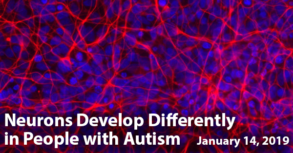
What’s New: A recent study explored the development of brain cells, or neurons, derived from iPSCs taken from people with autism spectrum disorder (ASD) as compared to those from their typically developing peers. The researchers took skin cell samples from 8 people with ASD and 5 people without ASD between the ages of 6 and 19. They transformed those cells into iPSCs, from which they coaxed neuronal cells.
In observing the development of the neuronal cells, the researchers found key differences in the cells that came from people with ASD, including:
- The cells grew to a bigger size with more complex branches.
- Networks of the cells behaved differently, with certain brain development processes beginning earlier.
- Those brain processes involved genes that have been associated with ASD.
Why it’s important: This study sheds light on the early development of ASD. Future studies could refine strategies for earlier diagnosis.
Image: Neuronal cells derived from induced pluripotent stem cells (National Center for Advancing Translational Sciences/National Institutes of Health)
Help me understand :
| Source(s) : |
| Tweet |
Children with ASD Show Patterned Sensory Behavior
By Chelsea Toledo, M.A. on February 28, 2018
Background: To date, autism is behaviorally diagnosed by trained clinicians using a comprehensive approach that includes systematic and structured observation of a child in the two areas: (1) Social interactions and Communications and (2) Restricted, repetitive patterns of behavior and interests. The range and degree of autism symptoms falls on a continuum, called the autism spectrum. Therefore, both children with severe deficits as well as those who are mildly affected are considered to have Autism Spectrum Disorder (ASD). However, little research has leveraged physiological markers or sensory information to classify ASD subtypes among children.
What’s New: A recent study explored a process for subtyping preschool aged children based on sensory processing patterns, along with social acumen, communication, motor skills, and adaptive behavior. The study employed a model called latent profile analysis, in which observable variables (such as scores on the Short Sensory Profile) are related to hidden variables (such as an underlying sensory subtype). After analyzing data from 400 children with ASD between the ages of 3 and 6, the researchers were able to define four subtypes:
- Sensorimotor – The largest subtype identified, this group had high scores in taste-smell sensitivity and sensory seeking, and was under-reactive to sensory stimuli such as heat or sound (hypo-responsivity). Compared to the other groups, this subtype was characterized by decreased language, social, and adaptive behavior skills. The average age of children in this group was 4 years old.
- Selective Complex – Like the Sensorimotor group, this subtype was characterized by sensory seeking, hyporesponsivity, and decreased language and social skills. However, developments varied among this group, with higher motor skills overall. The average age of children in this group was 4.5 years old.
- Perceptive-Adaptable – Children in this group showed similar patterns to those in the Selective Complex group – with high scores for motor skills and low scores for social acumen and language. However, the children in this group scored higher in those categories overall, as well as in adaptive behavior. The average age of children in this group was 4 years old.
- Vigilant-Engaged – The smallest subtype identified, this group had the hghest developmental skills, with high scores for language, adaptive behavior, and social acumen. They had increased sesntivity in auditory-visual stimuli, as well as taste and smell. This was the oldest group, with an average age of just over 4.5 years.
Why it’s important: This study explored a holistic method for sub-typing children with ASD – which could one day have implications on treatment and shed light on the disorder’s underlying cause. However, considering the groups differed significantly by age, these patterns should be researched further to define subtypes more concretely.
Help me understand :
| Source(s) : |
| Tweet |
Study Explores Brain Activity Related to Joint Attention
By Chelsea E. Toledo, M.A. on November 9, 2017

Background: Imagine a man and a woman having a conversation at a coffee shop while waiting for their orders. While they are making eye contact, the woman’s gaze shifts to the counter, where the man’s coffee has now appeared. The man then turns his attention to the drink, as well. This process is called joint attention – the shared focus on an object, cued by a verbal or non-verbal signal from one individual to another. Part of the communicative differences observed in Autism Spectrum Disorder (ASD) arises due to the lack of shared attention.
What’s new: On October 19, 2017, the journal Scientific Reports published a study that assessed the underlying brain activity of eleven 6 to 9 year old boys with high-functioning ASD in situations that called for joint attention. Over a period of six months, the researchers held weekly treatment sessions: the first half of the session was devoted to play-based activity, and the second half to administration of a tablet-based therapy.
To assess joint attention, the researchers recorded the children’s response to one therapist looking at the child and then gesturing to another therapist (responding joint attention), to having a story read by one therapist while another therapist acted the story out (initiating joint attention), and to other scenarios eliciting joint attention. In the second half of the session, they administered a tablet-based therapy game (called GOLIAH, based in the MICHELANGO framework that uses wearable technology to track brainwaves and eye movements).
The researchers collected data on brain activity and eye movements at the beginning and the end of the six-month study. They found that:
- Initiating and responding joint attention have both specialized and overlapping brain activity patterns
- There were changes in brain activity after treatment
- The trends in brain activity following treatment corresponded with modified eye movements
Why it’s important: This pilot study suggests that an approach integrating neuronal and eye-tracking data can provide a clearer picture of the brain’s activity during joint attention. Future studies with a larger sample of participants could shed light on the underlying reasons that joint attention differs in people in ASD – and could lay the groundwork for therapies targeting this skill.
Help me understand :
| Source(s) : |
| Tweet |
Tablet-Based Autism Research Reveals Social Insights
By Shana R. Spindler, PhD on October 24, 2017
Background: Babies are drawn to sights and sounds of the social world. A mother’s voice, a friendly face, the sound of laughter—all of these social experiences are important for a baby’s development. In toddlers and children with autism spectrum disorder (ASD), the amount of attention given to social matters is reduced. But to what extent, and at what ages, social interest differs is an active area of study. Newer technologies, such as tablet computers, have opened the door for novel ways to look at social preferences in children with ASD.
What’s new: On June 21, 2017, the journal Scientific Reports published a tablet-based study of children’s desire for social rewards. The researchers enrolled 63 children, ages 14 to 68 months, into the study (25 children with ASD and 38 typically developing children). Each child pressed buttons on a tablet screen that caused social or non-social pictures to appear. As the children played with the tablet, the researchers recorded the children’s social behaviors, such as smiles, vocalizations, and eye contact.
The children with ASD pressed buttons for non-social images at a higher rate than for social images, whereas the typically developing children lacked a preference for social versus non-social. For both groups of children, increased social image selection linked to an increase in social behaviors while playing.
Why it’s important: Many past studies of social interest in children with ASD relied on older children and passive viewing of images. This unique, tablet-based approach allowed researchers to test social reward-seeking in a younger group of children. The results are consistent with earlier findings that children with ASD favor non-social stimuli.
Help me understand :
| Source(s) : |
| Tweet |
Babies with Autism View Social Scenes Differently
By Chelsea E. Toledo, M.A. on September 25, 2017
Background: Humans are fundamentally social creatures. Starting in infancy, faces are critical for communicating; even before acquiring most basic skills, babies learn to “read” the emotions of others by watching their eyes and mouths. The brain's social pathway attaches meaning to social signals, motivates us to respond to social signals, and ultimately guides our social behavior.
What’s new: On July 20, 2017, the journal Nature published a study exploring the basis for how young children with and without autism spectrum disorder (ASD) processed such social information. The researchers tracked the eye movements of a total of 250 typically developing toddlers—including 82 monozygotic twins, 84 dizygotic twins, and 84 non-siblings—as they watched videos depicting social situations. They found distinct patterns in the eye tracking results based on the children’s genetics: compared to dizygotic twins, identical twins within the sample had very similar eye movements, especially when it came to their focus on eyes and mouths. Furthermore, the researchers compared eye-tracking data from 88 children with ASD. They found that the characteristic eye movement patterns in identical twins—the focus on eyes and mouths—were markedly reduced in children with ASD.
Why it’s important: In a series of well-designed experiments using eye-tracking measures, the authors of this study shed light on the genetic underpinnings of social information seeking in developing children. The researchers also provide evidence regarding how this trait may be altered in autism.
Help me understand :
| Source(s) : |
| Tweet |
Extra Stable Brain Activity Patterns Found in ASD
By Shana R. Spindler, PhD and Chelsea E. Toledo, M.A. on August 14, 2017
Background: Even at rest, the brain never really rests. Brain cells, or neurons, use chemicals to shoot information at lightening speed throughout the brain all the time. This brain activity occurs in special patterns that are related to the different functions of the brain. Scientists think that measuring patterns of brain activity during a resting state can offer clues about why some people have autism.
What’s new: On July 5, 2017, the journal Nature Communications released a study comparing resting brain activity in typically developing individuals versus their peers with high-functioning autism spectrum disorder (ASD). Researchers looked at brain imaging data taken in a resting state from a total of 50 male individuals between the ages of 18 and 39.
The researchers used a special type of mathematical formula to make brain activity look like peaks and valleys on a topographical map. They found that individuals with ASD had differently sized peaks and valleys as compared to their typically developing peers. This particular method of analyzing brain activity allowed autism identification—based solely on brain scans—with about 85% accuracy. The researchers also found a correlation between the peak and valley sizes and the severity of autism symptoms.
Why it’s important: The study is an important step forward in establishing ways to extract information from brain scans, which in this case was a functional magnetic resonance imaging (fMRI) scan. The findings of the study suggest a stability of brain activity pattern that differs between typically developing individuals and those with autism spectrum disorder. These differences may one day aid autism diagnosis or indicate severity along the spectrum, but additional investigation is required.
Help me understand :
| Source(s) : |
| Tweet |
Executive Functioning Linked to Autism Risk, Study Finds
By Chelsea E. Toledo, M.A. on August 25, 2016
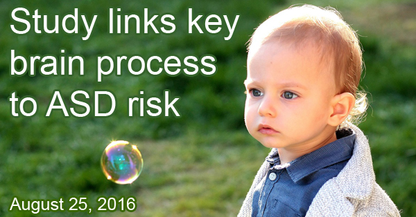
Background: Executive functioning refers to mental processes, such as planning, reasoning, and problem-solving. A key executive function is working memory, the attention to and monitoring of an ongoing task. While existing research has suggested a link between autism spectrum disorder (ASD) and deficits in executive functioning, the relationship has not been extensively studied.
What’s New: A recent study explored the relationship between executive functioning, specifically working memory, and motor skills in infants and toddlers at high and low risk for ASD. Using established motor skill assessments alongside a toy finding task, the researchers compared overall motor skills and executive functioning among a total of 262 children—first at 12 months, and then at 24 months. Children were assessed for ASD at the later time point, with 19 of the 186 high-risk children receiving a diagnosis. As a group, children at high risk of developing ASD (established by having a sibling with the disorder) showed less improvement in executive function than their low-risk peers over time. Those deficits were associated with poorer motor skills related to the suppression of actions.
The journal Frontiers in Psychology published the study on July 5, 2016.
Why it’s important: This study suggests that executive functioning and motor skills are affected in all children at high risk for ASD—even those who do not ultimately develop the disorder. Because the researchers looked at children before ASD diagnosis, the study provides the earliest look at executive function in children at high risk for autism.
Help me understand :
| Source(s) : |
| Tweet |
Motor Deficits with ASD Linked to Right Side of Brain
By Shana R. Spindler, PhD on August 10, 2016
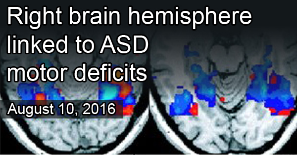
Background: At least 80% of children with autism spectrum disorder (ASD) have motor problems, according to recent estimates. Typical problems include delayed motor skills and trouble with coordination, such as kicking a ball or grasping small objects. In some cases, motor problems are apparent before other ASD symptoms.
What’s new: A new brain imaging study looked at motor control in the left and right sides of the brain in 8 to12 year old children with ASD. Researchers visualized brain activity, during a finger-tapping task, of 44 children with high-functioning autism and 80 typical, control children. Normally, regions in the left side of the brain specialize in language and motor functions. Previous work found that these regions are right-side dominant for language processing in children with ASD. Similarly, in this study researchers found brain regions that were right-side dominant for motor control too.
Why it’s important: This is the first study to look at left- and right-brain activity related to motor functions in children with ASD. The finding that some motor control is shifted to the right side of the brain in ASD is important given the potential for brain imaging to provide markers for early diagnosis.
This work was published on July 14, 2016, in the journal Molecular Autism.
Help me understand :
| Source(s) : |
| Tweet |
ASD Traits with Anxiety Linked to Reward Areas in Brain
By Shana R. Spindler, Ph.D. on July 26, 2016
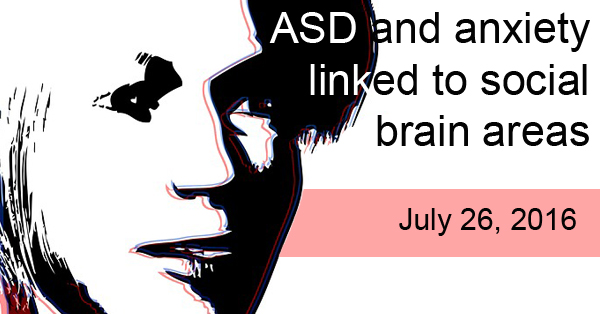
Background: The brain has specific areas that help you think about rewards, such as reward anticipation or outcomes from receiving an award. This is called reward processing. Previous studies have shown that reward processing is different in people with autism spectrum disorder (ASD). But it’s not clear if this is due to ASD alone, or conditions that sometimes occur with ASD. For example, nearly 40% of people with ASD also have anxiety.
What’s new: A new study published on June 28, 2016, suggests that ASD and anxiety share some regions of the brain associated with reward processing. Researchers looked at brain activity from 70 teenagers with high ASD traits and 1402 teenagers with low ASD traits. They found a unique pattern of activation in reward-related areas of the brain in those with high ASD traits combined with anxiety. But not all neuroimaging findings were shared when ASD traits and anxiety were present on their own.
Why it’s important: The results of this study point to patterns of brain activity related to reward processing that might link ASD with anxiety. If reproducible, these findings may allow doctors to use the patterns seen during neuroimaging to help diagnose specific subgroups of ASD, such as ASD with co-occurring conditions.
Help me understand :
| Source(s) : |
| Tweet |
Newborns at Risk for Autism Differ in Social Preferences
By Shana R. Spindler, Ph.D. on June 9, 2016
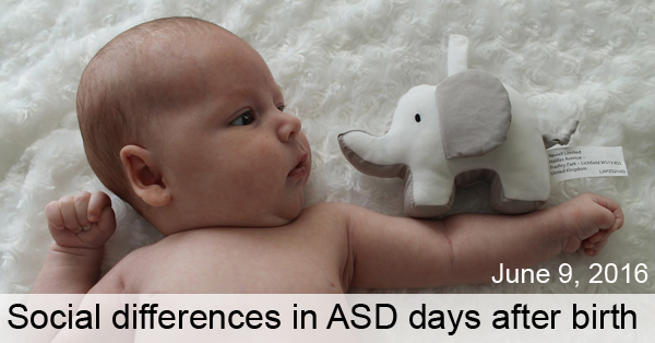
Background: Newborn babies pay close attention to relevant social cues, such as a mother’s face. Some researchers hypothesize that the social preferences typically seen in infancy are impaired in autism, leading to an underdevelopment of “social brain” networks.
What’s new: On May 20, 2016, Scientific Reports published a study that examined social preferences in newborns at high risk for autism. The researchers showed social and non-social images to 13 newborns with high risk of autism and 16 newborns with low risk of autism—all between 6 and 10 days old. Researchers scored each newborn on how much he or she looked at the social versus non-social images. According to the report, the newborns at high risk for autism looked significantly more at the non-social images than the low-risk infants did.
Why it’s important: This is the first study to show that babies at high risk for autism show diminished social attention as early as 6 days after birth. Future research could include a larger number of infants and follow up to see if the high-risk infants receive an Autism Spectrum Disorder diagnosis.
Help me understand :
| Source(s) : |
| Tweet |
New Evidence Contradicts Popular Autism Theory
By Anjali Sarkar, PhD on June 2, 2016

Background: In neuroscience, a “simple task” involves activity in one or a few brain regions while a “complex task” involves the coordination of multiple regions in the brain. According to some neuroscientists, autism is marked by superior simple task performance but poor complex task performance.
This observation has led to a theory that diverse areas of brain activity fail to function as a whole in Autism Spectrum Disorder (ASD), leading to the social and behavioral symptoms common to the disorder. A specialized version of this theory focuses on problems with multisensory integration, how the brain combines information from all five senses. For example, when children with ASD are around noisy distractions, they tend to perform poorly on tasks that require attention, according to previous research.
What’s new: A recent study shows that children with ASD have intact multisensory integration. The researchers found that high-functioning adolescents with ASD performed as well as typical adolescents on multisensory tasks that combined visual sensory inputs from the eye and balance sensory inputs from the inner ear.
Why it’s important: The results of this study contradict the popular notions of defective multisensory integration in individuals with ASD. Instead, the study reveals an increased sensitivity to sensory inputs, likely due to problems with lesser understanding of the world. Because participants of this study are from the high-functioning end of the autism spectrum, future studies could test if children across the spectrum have proper multisensory integration.
Help me understand :
| Source(s) : |
| Tweet |
Non-invasive Brain Monitoring, a Potential ASD Therapy
By Chelsea E. Toledo, M.A. on April 19, 2016
Background: Among the treatments being considered for autism spectrum disorder (ASD), neurofeedback shows an indication of brain activity in real time, which helps people learn to regulate how their brains are functioning. The most common form of neurofeedback is analyzed using electroencephalography (EEG), which involves placement of electrodes on the scalp to measure changes in electrical activity within the brain. This process is frequently used to diagnose epilepsy.
What’s new: On January 14, 2016, the journal, Frontiers in Human Neuroscience published a study exploring EEG’s potential as a treatment for ASD, with a focus on the relationship between different frequency bands generated as a result of recording specific waveform oscillations in the brain’s activity. Researchers used the technology to monitor the brain activity of 18 participants with high-functioning ASD. After 18 weekly EEG sessions, they found a reduction in EEG characteristics associated with ASD in the brain’s prefrontal cortex – specifically those related to focused attention. They also saw improvement in lethargy and social withdrawal, as measured in an established questionnaire that was administered to parents at the beginning and end of the study.
Why it’s important: This study suggests that using neurofeedback from EEG analysis is a potential therapy for high-functioning children with ASD. Future research could hone in on specific techniques that compare relationships between frequency bands for the most effective method of treatment.
Help me understand :
| Source(s) : |
| Tweet |
Language Delay in ASD Linked to Brain Symmetry
By Chelsea E. Toledo, M.A. on March 29, 2016
Background: Autism spectrum disorder (ASD) encompasses a broad array of conditions characterized by differences in socialization, communication, and behavior. One area where people with ASD can differ widely from one another is in language acquisition. Some individuals face severe delays in speech while others progress more typically.
What’s New: The January 2016 issue of Human Brain Mapping featured a study that explored the symmetry between brain hemispheres in individuals with or without language delay – defined as uttering one’s first words after 24 months of age or first phrases after 33 months of age. The researchers performed structural magnetic resonance imaging (MRI) on 136 right-handed men between the ages of 18 and 43 (67 with ASD and 69 with typical development). Participants in the ASD group had reduced asymmetry between several brain areas across the left and right sides of the brain, which was most pronounced in those with language delay and/or abnormal social functioning.
Why it’s important: This structural asymmetry of the brain could eventually help clinicians diagnose and understand specific subgroups of ASD. Future research could examine the brain symmetry of women and/or left-handed individuals with ASD.
Help me understand :
| Source(s) : |
| Tweet |
Study Probes Normalized Symptoms of Autism
By Chelsea E. Toledo, M.A. on March 22, 2016
Background: Autism spectrum disorder (ASD) is generally characterized by differences in socialization, communication, and behavior. Research has shown differences in brain activity underlying these traits. However, certain individuals have been shown to “grow out” of ASD symptoms, with social, communicative, and behavioral traits mirroring those of their typically developing peers. Little is known about this phenomenon—do individuals experience suppression of atypical brain activity, or does the brain establish compensatory mechanisms?
What’s New: On December 2, 2015, the journal NeuroImage: Clinical published a study exploring the underlying brain activity of people with ASD whose symptoms became normalized later in life. The researchers administered a sentence comprehension task while performing brain scans on 59 participants between the ages of 8 and 21 (23 with high-functioning ASD, 20 with typical development, and 16 who no longer met the criteria for an ASD diagnosis). They found similar activity between the ASD and normalized ASD groups, with enhanced compensatory brain activation in the group with normalized symptoms.
Why it’s important: This study suggests that people with ASD do not “grow out” of the disorder, as their brain activity more closely mirrors that of people with ASD than those with typical development when processing language. Instead, the brain may compensate by increasing activity in language processing areas of the brain. Future studies could investigate patterns of brain activity when completing other types of tasks, and additional research may reveal the role of different types of therapy in ameliorating ASD symptoms.
Help me understand :
| Source(s) : |
| Tweet |
Different Visual Processing Patterns Observed in Autism
By Chelsea E. Toledo, M.A. on February 23, 2016
Background: On a visual plane, the oblique axis runs at a 45 degree angle relative to the horizontal and vertical axes. For typically developing individuals, it is easier to perceive patterns along the vertical and horizontal axes than those along the oblique axis—a phenomenon known as the “oblique effect.”
What’s New: On January 21, 2016, the journal Frontiers in Neuroscience published a study exploring how people with autism spectrum disorder (ASD) differed from their typically developing peers on visual processing tasks. The researchers administered the same task to 64 boys between the ages of seven and fifteen, 26 of whom had ASD. Each participant was tasked with looking at a striped object tilted at various angles, and to determine its position relative to the vertical and oblique axis. The group with typical development perceived the relationship of objects to the vertical axis much better than the relationship of objects to the oblique axis – a pattern not observed in the group with ASD.
Why it’s important: This study suggests that children with ASD have impaired visual processing along a vertical axis, but not along an oblique axis. Future studies could pinpoint the neurological basis of this difference, revealing clues about the underpinnings of ASD.
Help me understand :
| Source(s) : |
| Tweet |
Visual Study Links Neurotransmitter to Autism
By Chelsea E. Toledo, M.A. on February 9, 2016
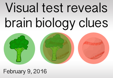
Background: Neurotransmitters are chemicals that send messages within the brain, contributing to a range of every day functions. There are seven primary neurotransmitters in humans: acetylcholine, dopamine, serotonin, norepinephrine, epinephrine, gamma-amino butyric acid (GABA), and histamine. GABA is the major inhibitory neurotransmitter in both the brain and spinal cord – helping to suppress excess stimuli. Previous studies suggest that Autism Spectrum Disorder (ASD) may stem from an imbalance between excitatory and inhibitory signals in the brain.
What’s New: On January 11, 2016, the journal Current Biology published a study exploring the relationship between GABA and the behavioral symptoms of people with ASD. The researchers administered a visual test to 20 individuals with autism and 21 with typical development, showing images in both eyes and testing how well the participants suppressed one image to focus on another—typically, the brain alternates focus between each eye and the singular mixed image. The researchers found that the participants with ASD were significantly slower to switch their focus between the images. The team then used a specialized brain imaging technique on the participants and found a correlation between the visual task performance and GABA activity in the brain.
Why it’s important: The ability to switch focus between two visual cues is thought to rely on the brain’s ability to control the inhibition and excitation of neurons, making this a good model to study inhibitory signals in the brain. Future studies could delve deeper into this relationship, determining the precise role of GABA inactivity in ASD.
Help me understand :
| Source(s) : |
| Tweet |
Children of Blind Parents Reveal Clues About Eye Contact
By Chelsea E. Toledo, M.A. on January 12, 2016

Background: Children with Autism Spectrum Disorder (ASD) have been known to avoid eye contact. Studies have demonstrated that children who eventually receive a diagnosis can display differences in eye contact as early as six months of age. Researchers have hypothesized that this tendency could impact the development of areas in the brain related to social behaviors.
What’s New: On November 19, 2015, the journal Current Biology published a study of sighted children born to blind parents. The researchers used eye-tracking technology to explore how 14 infants between the ages of 6 and 10 months engaged with adult faces, repeating the experiment 6 months later. When compared to a group of infants born to sighted parents, children born to blind parents paid less attention to adults’ eyes – as seen in similarly aged children with ASD. However, the children of blind parents demonstrated otherwise typical development, and even excelled at visual attention and memory.
Why it’s important: This study shows that typical children adjust their behavior as they adapt to their environments and social cues. If a parent uses eye movements, rather than words and gestures, to signal important objects, then a child will focus more on the parent’s eyes. Future studies could address if core ASD traits stem from a child’s inability to adapt to his or her early environment appropriately.
Help me understand :
| Source(s) : |
| Tweet |
Brain Organization Ages Differently in Autism
By Chelsea E. Toledo, M.A. on November 24, 2015
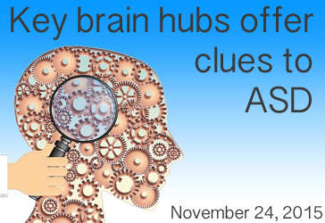
Background: The human brain is comprised of an intricate network of interconnected regions. Of the hundreds of regions in the human brain, there exists highly connected central regions--or hubs--that link sparsely populated peripheral regions of the brain. Known as the “rich club,” these areas are thought to play key role in integrating information across the brain. Research has suggested that their connectivity may be different in people with autism spectrum disorder (ASD).
What’s New: On November 5, 2015, the journal Scientific Reports explored how organization of the rich club hubs differed in people with ASD versus those with typical development as they aged. The researchers compared brain connectivity data of children between the ages of 9 and 13 to that of a group of adolescents between the ages of 13 and 18. The results of their analysis indicated that organization of rich club hubs in the brain increased as the 36 typically developing children and adolescents aged, but that the same progress wasn’t observed in the 45 children and adolescents with ASD.
Why it’s important: This study adds to the findings suggesting that rich club organization underlies cognition, and that dysfunction in that organization could play a role in certain neurodevelopmental disorders. Future studies could illuminate the precise role of rich club organization and pinpoint how puberty affects its development.
Help me understand :
| Source(s) : |
| Tweet |
Neural Basis of Noise Overload in ASD Explained
By Shana R. Spindler, PhD on November 17, 2015
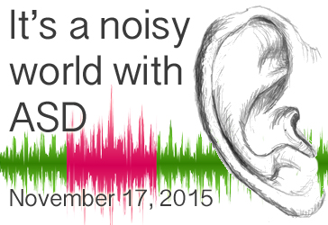
Background: About 90 percent of people with Autism Spectrum Disorder (ASD) experience some form of sensory-related symptoms, such as over or under sensitivity to sights, sounds, and even tastes (reviewed in Geschwind, 2009). Researchers have hypothesized that an abnormal adaptation to sensory stimuli may underlie the social and communication difficulties seen in ASD.
What’s new: On November 5, 2015, Scientific Reports published a study examining loudness adaptation in 20 adults with ASD and 20 neurotypical individuals. The researchers found that adults with ASD were slower and less able to adapt to a continuous, low-level sound than neurotypical adults.
In the study, the participants listened to a soft, constant sound for several minutes and recorded the perceived volume of the sound at specific time intervals. While the neurotypical individuals said the sound decreased by about 50 percent in volume over time—even though the volume of the sound was unchanged—adults with ASD perceived a mere 20 percent reduction. In contrast, adaptation to loud sounds with intermittent disruptions was similar between neurotypical adults and those with ASD.
Why it’s important: The adaptation to constant, quiet sounds is thought to involve a different part of the brain than adaptation to loud, interrupted sounds—think of the hum of a refrigerator versus a fire alarm. Therefore, this is the first study to show that noise overload in ASD may stem from a neural inability to adapt to certain sound types. Interestingly, for individuals with ASD who reported using coping strategies, their adaptive responses were closer to neurotypical individuals.
Help me understand :
| Source(s) : |
| Tweet |
How People with ASD See the World Differently
By Chelsea E. Toledo, M.A. on November 10, 2015

Background: Autism spectrum disorder (ASD) affects three major and inter-related areas: communication, social interaction, and behavior. Research has shown that people with the disorder read faces and other social cues differently than their typically developing peers, which could contribute to the observed behavioral symptoms in ASD.
What’s New: On November 4, 2015, the journal Neuron published a study exploring how general gaze – not just at faces – may differ in individuals with ASD. The researchers showed 700 naturalistic images capturing common daily scenarios to 20 people with ASD and average IQ. Using an eye-tracking device to follow their gaze, they found that, when compared to 19 peers with typical development, the ASD group spent more time focused on the center of an image, even if there was no object in the image’s center. They also found that the ASD group took longer to focus on faces in the images, but were quicker than their typically developing peers to focus on mechanical objects.
Why it’s important: This study suggests that differences in gaze observed in people with ASD is not limited to reading faces, but represents a larger perceptive difference. Larger studies are needed to further define these differences in atypical visual processing in ASD. The eye tracking methodology holds much promise as a non-behavioral early ASD diagnostic marker.
Help me understand :
| Source(s) : |
| Tweet |
Face-Reading Study Supports “Extreme Male” Theory
By Chelsea E. Toledo, M.A. on September 9, 2015
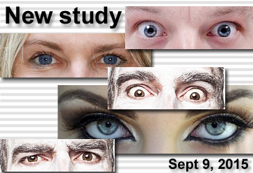
Background: Autism spectrum disorder (ASD) is four times more common in boys than in girls. That trend has led some researchers to subscribe to the “extreme male theory” – that the brain of a person with the disorder is essentially an extreme version of male brain in terms of its structure and function.
What’s new: On August 27, 2015, the journal PLoS ONE published a study exploring sex differences when it comes to interpreting facial expressions. The researchers administered an existing online test – called “Reading the Mind in the Eyes” – to a total of 715 adults at an average age of 39. The scores of 152 men and 168 women with typical development followed the previously established trend: women scored better at the task of choosing the correct emotion to match a given facial expression. However, the scores of the 178 men and 217 women with ASD were nearly identical – and much lower across the board than those of their typically developing peers.
Why it’s important: While the “Reading the Mind in the Eyes” test has been used in hundreds of studies since its development in 1997, this is the largest study to administer the test to people with ASD. Future studies could reveal more about the underlying causes of ASD by delving deeper into the sex-common and sex-specific features of ASD.
Help me understand :
| Source(s) : |
| Tweet |
Regressive ASD Linked to Temporal Lobe Activity
By Chelsea E. Toledo, M.A. on September 1, 2015
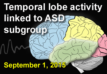
Background: The temporal lobe, one of the four major parts of the brain’s cerebral cortex, aids in processing sound, categorizing objects, and consolidating memories. It also plays a role in visual processing, emotions, and language. Researchers first began to suspect a relationship between temporal lobe abnormalities and autism spectrum disorder (ASD) in 1975. (see second Pubmed link below)
What’s New: On July 30, 2015, the journal European Child & Adolescent Psychiatry published a study investigating if irregular electrical brain activity – an indicator of epilepsy – could predict any specific feature of ASD. The researchers analyzed the results of brain scans from 220 children and young adults, 71 with ASD and 146 with other developmental disorders. After performing brain measurements on a subgroup of participants, they found a correlation between abnormal electrical brain activity, relatively large head size, and loss of previously acquired developmental skills, known as regression. The relationship between regression and abnormal electrical brain activity was the strongest when that activity took place in the temporal lobe.
Why it’s important: The results of this study support the possibility that irregular electrical activity in distinct brain areas may predict or help define ASD subgroups.
Help me understand :
| Source(s) : |
| Tweet |
Level of Hormone Predicts Theory of Mind Ability in ASD
By Shana R. Spindler, Ph.D. on August 13, 2015
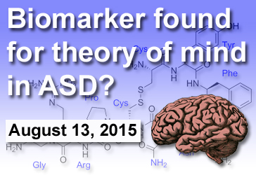
Background: Difficulty with social functions is a defining characteristic of Autism Spectrum Disorder (ASD). Hormones in the brain, such as oxytocin and arginine vasopressin, play an important role in regulating social behaviors in humans and other mammals. While several studies have focused on the potential role for oxytocin in the diagnosis and treatment of ASD, few investigations have centered on arginine vasopressin.
What’s new: On July 22, 2015, the online journal PLOS ONE published a study examining the relationship between blood levels of arginine vasopressin (AVP) and social abilities in those with ASD, their siblings, and neurotypical controls. The researchers first confirmed that blood levels of AVP predicted brain levels of the hormone. Next, they found that ASD individuals with lower blood levels of AVP have decreased Theory of Mind—the ability to interpret another’s intensions and emotions. This correlation is specific only to Theory of Mind ability, as AVP blood levels did not predict other social function scores.
Why it’s important: ASD is a heterogeneous condition—individuals along the spectrum have varying degrees and combinations of symptoms. Consequently, there is individual variability in Theory of Mind. Having a biomarker for this nearly universal feature of ASD will be important for diagnosis and treatment selection. This study suggests that the blood level of arginine vasopressin could be a biomarker for theory of mind ability in individuals with autism.
Help me understand :
| Source(s) : |
| Tweet |
Size of Brain Area May Predict ASD in Infants
By Chelsea E. Toledo, M.A. on July 1, 2015
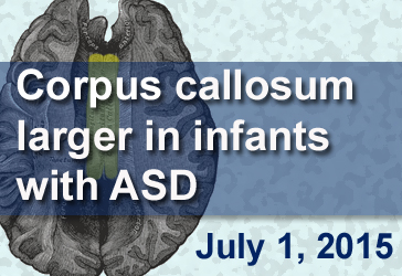
Background: “White matter” refers to the parts of the brain that transmit signals between brain regions. The brain’s largest white matter connection, the corpus callosum, is a thick bundle of fibers connecting the brain’s hemispheres and facilitating communication between them. Numerous studies have shown that the corpus callosum is smaller in adults and children with autism spectrum disorder (ASD) than in their typically developing counterparts.
What’s New: On May 3, 2015, the journal Brain published the first study focusing on the corpus callosum in infants at risk of developing ASD. The researchers scanned the brains of 378 children—270 with older siblings diagnosed with ASD—at 6, 12, and then 24 months of age. Fifty-seven of those children fit the ASD profile at 24 months of age. In those children, the corpus callosum was much thicker—especially at the 6- and 12-month time points, with differences diminishing as children approached 2 years of age. The finding held even when the researchers controlled for brain size, which studies have shown is larger in infants with ASD.
Why it’s important: This is the first study to examine how the size of the corpus callosum during infancy relates to ASD diagnosis. Future research could examine the biological mechanisms that lead to changes in relative corpus callosum size from infancy to child- and adulthood in those with autism. Several lines of evidence point to abnormalities in the way neuron growth and insulation is managed in the brain.
Help me understand :
| Source(s) : |
| Tweet |
Study Links Autistic Traits to Adult Disorders
By Chelsea E. Toledo, M.A. on May 12, 2015
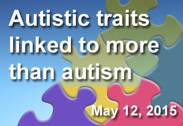
Background: Autism Spectrum Disorder (ASD) presents itself as differences in communication, social interaction, and behavior. Research has shown that these traits often appear in children with psychiatric conditions such as depression and bipolar disorder. However, fewer studies have evaluated whether adults affected by psychiatric disorders also exhibit autistic-like traits.
What’s New: On April 2, 2015, the online journal PLOS ONE published a study exploring the prevalence of autistic-like traits in adults with psychiatric disorders. The researchers applied a series of established psychological and behavioral screenings to a total of 290 individuals between the ages of 25 and 59—125 with clinical depression, 56 with bipolar disorder, 44 with schizophrenia, and 65 with no psychiatric diagnosis. They found that a significant portion of the individuals with psychiatric disorders—with the exception of depression in remission—exhibited autistic-like traits like restrictive, repetitive behaviors. The prevalence of these ranged from 46 percent in those with ongoing depression to 61 percent in those with schizophrenia—as compared to the 14 percent of individuals with no psychiatric diagnosis who demonstrated autistic-like traits.
Why it’s important: This is the first major study to examine the link between adult psychiatric conditions and the traits observed in ASD. Because those traits were so strongly associated with adult bipolar disorder and schizophrenia, the findings could inform screening and treatment for those patient populations.
Help me understand :
| Source(s) : |
| Tweet |
Study Links Brain Differences to Language Delay
By Chelsea E. Toledo, M.A. on May 4, 2015
Background: Autism Spectrum Disorder (ASD) varies widely in its presentation. While some individuals on the spectrum have no history of language delay, others experience extreme differences in behavior, social skills, and communication compared to peers. Several studies have addressed the brain differences between individuals with ASD and those with typical development, but little research has focused on how brain development might differ for those with varying presentations of the disorder.
What’s new: On March 3, 2015, the journal Autism Research published a study exploring brain differences in adults with ASD to determine whether those who had experienced language delay were neurologically different from those who had not. Every participant had an IQ in the normal range. The researchers found that the 27 individuals who had experienced language delay had thinner layers in a specific brain region than the 37 individuals with typical language development. The thickness of that brain region, commonly associated with cognition, correlated with higher verbal IQ scores.
Why it’s important: This is the first study to link verbal IQ to differences in specific regions of the brain’s outer layers. This work could help researchers understand the underlying factors leading to the variation in different cases of ASD.
Help me understand :
| Source(s) : |
| Tweet |
Study Pinpoints Differences in Autistic Brain
By Chelsea E. Toledo, M.A. on March 31, 2015
Background: Autism Spectrum Disorder (ASD) is characterized by differences in social interaction, communication, and behavior. Researchers have hypothesized that the functioning of—and communication between—related brain regions is atypical in people affected by the disorder. Their studies have largely focused on select areas of the brain, as opposed to observing them within the context of the entire brain system.
What’s new: On March 20, 2015, the journal Brain published a large study providing a whole-brain perspective of ASD versus typical development. The researchers used a grid-based technique to analyze the entire brain at once with data from existing functional Magnetic Resonance Imaging (fMRI) scans of the resting brain. They found that, in comparison to 509 typically developing individuals, the 419 participants with ASD showed differences in 20 key areas.
Why it’s important: The observed differences were in areas of the brain active in processing facial expressions, sense of self, and theory of mind—the ability to discern and predict other’s mental states. Future studies could leverage this technique to learn more about brain function in obsessive compulsive disorder, attention deficit hyperactive disorder, and schizophrenia.
Help me understand :
| Source(s) : |
| Tweet |
Brain Awareness Week 2015
By Shana R. Spindler, Ph.D. on March 16, 2015
March 16-22 marks Brain Awareness Week 2015, a worldwide campaign coordinated by the Dana Alliance for Brain Initiatives and the Society for Neuroscience to promote the progress and benefits of brain research. Hundreds of international events are underway in celebration of this unique week.
Autism Reading Room offers extensive coverage of brain biology, from development to function, in our Brain Biology area. Check it out!
Help me understand :
| Source(s) : |
| Tweet |
Scientists Piece Together Chemical Imbalances in ASD
By Chelsea E. Toledo, M.A. on December 11, 2014
Background: The chemicals serotonin and melatonin transmit important signals between brain cells. Serotonin helps to regulate mood, but it also converts into melatonin, an important regulator of sleep cycles. Previous studies have found separately that individuals with autism spectrum disorder (ASD) have elevated levels of serotonin and diminished levels of melatonin. That chemical imbalance is a suspected contributor to the behavioral and sleep issues observed in people with ASD.
What’s new: On November 11, 2014, the journal Translational Psychiatry published a study examining both chemicals—as well as N-acetylserotonin (NAS), the chemical serotonin becomes in the process of converting to melatonin. The researchers monitored levels of serotonin, NAS, and melatonin in the blood, platelets, and plasma, respectively, of 278 patients with ASD. When compared to 506 relatives unaffected by the disorder—as well as a control group of 416 people matched by age and sex—the group with ASD experienced significant disruption in the conversion of serotonin to NAS to melatonin. This disturbance was evidenced by high levels of serotonin and NAS and low levels of melatonin—with effects appearing most prominently in individuals with ASD and, to a lesser extent, in their blood relatives.
Why it’s important: This study sheds light on the relationship between serotonin, melatonin, and the intermediate chemical NAS as they relate to ASD. Future research can refine tests for disruptions in the level of these chemicals as biomarkers for the disorder. In addition, this study supports the possibility of melatonin as a therapeutic sleep aid for affected patients.
Help me understand :
| Source(s) : |
| Tweet |
New Hypothesis of ASD as a Disorder of Prediction
By Chelsea E. Toledo, M.A. on October 17, 2014
Background: Autism spectrum disorder (ASD) is characterized by difficulties with social communication and tendencies to engage in restrictive, repetitive behaviors. While those traits have been widely recognized, research has yet to determine whether they share a common, underlying cause.
What’s New: On October 6, the Proceedings of the National Academy of Sciences published a study describing a new hypothesis: that ASD symptoms stem from an inability to predict what’s coming next. The researchers suggested that people with the disorder experience events “as if by magic”—with the cognitive systems for linking one happening in their environment to another somehow compromised. While their hypothesis doesn’t explain every aspect of the autism profile, the researchers asserted their belief that predictive impairment underlay two of the most salient and seemingly disparate traits: difficulties with social communication and tendencies to engage in restrictive, repetitive behaviors. For example, the same predictive impairment that inspires an individual to line up objects for comfort could also be at play when that individual experiences difficulty understanding intention of others. Finally, a reduction in motor anticipation could lead to atypical gesture and posture often observed in ASD children.
Why it’s important: This study provides a theoretical framework to explain various features of ASD. With this approach, researchers might be able to identify structures in the brain with differences ascribed to impaired predictive abilities.
Help me understand :
| Source(s) : |
| Tweet |
ASD Subgroup Linked to Brain Volume
By Shana R. Spindler, PhD on October 7, 2014
Background: Autism Spectrum Disorder (ASD) features a diverse set of social and behavioral deficits, language abilities, and skill sets. Identifying unique subgroups in the ASD population is a major goal within the autism research community. Given that individuals within a subgroup may share causal risk factors, a subgroup diagnosis could enable targeted therapy options.
What’s new: Researchers in the United Kingdom have identified a subgroup of ASD based on language delay in childhood. The team used a technique called voxel-based morphometry to measure brain volumes in 80 adult men with ASD. Of the participants, 38 experienced childhood language delay with 42 reporting normal language development. Individuals with delayed language had an overall larger volume of brain grey matter than those with typical language histories. When the team measured specific brain areas, they found several clusters of decreased volume in those with language delay.
Why it’s important: Last year, the Diagnostic and Statistical Manual of Mental Disorders-Fifth Edition (DSM-5) grouped anyone along the autism spectrum into a single diagnosis of ASD. This study highlights the need to assess individual traits within individuals on the spectrum as we work towards understanding the underlying biology of ASD subgroups.
Help me understand :
| Source(s) : |
| Tweet |
ASD Diagnosis Increased with Additive Risk Factors
By Chelsea E. Toledo, M.A. on September 23, 2014
Background: Some researchers hypothesize that autism stems from a single causal event that initiates a cascade of cognitive and developmental delays. In contrast, others hypothesize that autism is the cumulative result of several co-existent risk factors. Studies have shown that disparities in social attention in children as young as 10 months old can predict whether those children later receive an ASD diagnosis, especially if they have an older sibling with ASD. Separate research has evaluated non-social attention—for example, how quickly a child can shift focus from one stimulus to another—and found differences in children as young as 6 months old, depending on their level of risk for developing ASD. By utilizing social and non-social attention tasks, researchers hope to learn about autism’s initial causes.
What’s new: The July 2014 issue of Developmental Science included a study assessing both social and non-social attention in children at 13 months of age. Using data from previous studies evaluating the two factors separately, the researchers combined data collected from a total of 145 children who had either undergone a task to follow the gaze of a person shown on a screen or the task to shift focus from one stimulus shown on a screen to another. They found that disparities in social and non-social attention had cumulative effects in predicting an ASD diagnosis—that is, infants who followed the gaze of the person on the screen for short periods of time and who also took long periods of time to shift from one task to another had a higher chance of developing ASD than those who scored better on each task.
Why it’s important: This is the first study to formally test the relationship between multiple factors (i.e. social and non-social attention) as they relate to the development of ASD. This research paves the way for future studies to more conclusively determine whether ASD develops due to cumulative risk, as this study suggests, or from a single causal factor.
Help me understand :
| Source(s) : |
| Tweet |
Too Many Neurons Partnered Up in Autism
By Ishita Das, PhD on September 19, 2014
Background: Soon after birth, the brain eliminates a large number of neural connections—a process called pruning. Researchers believe this fine-tuning affects connections that are weaker or in excess. Pruning proceeds alongside the earliest phases of learning. It likely helps the brain develop the sophisticated circuitry needed to respond to the environment. Researchers are investigating if pruning abnormalities underlie Autism Spectrum Disorder (ASD).
What’s new: A new study suggests that children and adolescents with ASD fail to eliminate excess neural connections after birth. New York researchers report that neurons in the brain’s temporal lobe contain more tree-like protrusions, known as spines, in adolescents with ASD compared to controls. This difference is discernable in brain samples from children with ASD as well. These spines receive input from other neurons. The temporal lobe is associated with language and speech perception, auditory and visual processing, and has been implicated in ASD etiology.
Using a mouse model, the group found that knocking out a pathway responsible for degrading parts of the cell reduced spine pruning and led to a significant increase in spine density in adolescent mice. Furthermore, using additional mouse models, the team connected spine density changes to deficiencies in social behavior and social interaction in the mice. Treatment with a drug, rapamycin, alleviated many of the social deficiencies seen in an ASD mouse model.
Why it’s important: This is the first study to show that a specific biological pathway is important for spine pruning, which this study implicates in ASD. Connecting these physical changes in the brain with behavioral features in mice is a major accomplishment in behavioral neuroscience.
Help me understand :
| Source(s) : |
| Tweet |
Making Connections: Target Practice
By Chelsea E. Toledo, M.A. on August 13, 2014
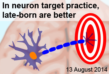
Background: An adult’s brain contains billions of nerve cells, called neurons, intricately connected to carry the signals controlling our senses and behaviors. During embryonic development, the nervous system begins to take shape around the fifth week of clinical gestation. Very little research has demonstrated how neurons of this early nervous system establish proper connections in the healthy brain – and how mistakes may lead to neurological disorders.
What’s New: On July 31, 2014, a study exploring the birth order of nerve cells appeared in the journal Cell Reports online ahead of print. The researchers focused on the cells connecting the eyes to the brain—called retinal ganglion cells (RGCs)—in mice. They found that early-born RGCs sampled many sites before establishing their final connection with other brain cells. By contrast, later-born RGCs were selective from the start in establishing neurological connections.
Why it’s important: This study suggests that the order in which brain cells form is important to how neurons establish proper neurological configuration. Future studies could determine whether the neurological dysfunction observed in disorders like autism are linked to differences in brain cells’ birth order.
Help me understand :
| Source(s) : |
| Tweet |
Empathy For Others in Pain Present in ASD
By Shana R. Spindler, Ph.D. on January 23, 2014
Background: Empathy is the ability to understand what another person is feeling. The lack of this trait was thought to underlie some of the social problems present in people with Autism Spectrum Disorder (ASD). However, few studies have addressed the brain activity of those with ASD in situations where empathy would be likely.
What’s new: On January 14, 2014, the online journal Translational Psychiatry published a study that suggests people with ASD maintain the neural ability to process empathy, at least when seeing others in pain. Researchers used functional magnetic resonance imaging to examine the spontaneous brain activity of 38 adolescents and adults with high-functioning ASD and 35 age-matched controls as they watched two-second videos of people with shoulder pain.
Contrary to popular belief, the individuals with ASD showed no significant difference in spontaneous brain activity upon seeing someone in pain as compared to the control group. Both groups had activity in brain areas involving emotional arousal and understanding. At a lower statistical threshold, the group with ASD appeared to have increased activity in brain areas likely used for situation assessment.
Why it’s important: The researchers suggest that empathy remains intact in ASD, while the perceived lack of caring links to attempts to calm personal distress by reassessing the situation. The authors of the study note that pain is a very basic social cue and that future studies should look at additional situations where empathy is used.
Help me understand :
| Source(s) : |
| Tweet |
It’s Brain Awareness Week!
By Shana R. Spindler, PhD on March 13, 2013
March 11-17 marks Brain Awareness Week 2013, a worldwide campaign coordinated by the Dana Alliance for Brain Initiatives and the Society for Neuroscience to promote the progress and benefits of brain research. Over 900 international events are underway in celebration of this unique week.
Expand your knowledge of the brain by investigating the “Brain Biology” corner of Autism Reading Room!
Help me understand :
| Source(s) : |
| Tweet |
Deep Brain Stimulation Improves Autism Symptoms
By Chelsea E. Toledo, M.A. on February 7, 2013
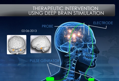
Background: Studies have shown that individuals with Autism Spectrum Disorder (ASD) have up to a 20 percent chance of injuring themselves. In the 1960s and 1970s, interventions were shown to improve epilepsy-related aggression when doctors targeted the amygdala, a brain region associated with emotional learning.
What’s New: In the January 21, 2013, issue of the journal Frontiers in Human Neuroscience, researchers published a study exploring the hypothesis that stimulating the amygdala would alleviate self-injurious behavior (SIB) related to ASD. They surgically implanted electrodes in the amygdala of a 13-year-old boy suffering from Kanner’s autism, also referred to as early infantile autism, and severe SIB. When the electrodes placed in the amygdala’s basolateral section were stimulated, the boy made significant strides in overcoming SIB as well as the emotional, social, and cognitive deficits related to autism.
Why it’s important: This study, the first to demonstrate such results in a patient, suggests that the amygdala—especially its basolateral section—is important for understanding behaviors related to ASD, and could provide a potential target in its treatment.
Help me understand :
| Source(s) : |
| Tweet |
Sex-Related Social Differences Amplified in ASD
By Chelsea E. Toledo, M.A. on January 30, 2013
Background: Autism Spectrum Disorder (ASD) appears about four times more frequently in males than in females. In typically developing individuals, women often score better than men on tests for social cognition and empathy—traits for which autistic individuals often show deficits. Therefore, some researchers have suggested that ASD could be a disorder in which male social patterns in the brain are exacerbated, known as the “extreme male brain theory” of autism.
What’s New: On December 26, 2012, the online journal PLoS ONE published a study examining whether women and men have markedly different social brain functions and whether those sex-related differences are prevalent in individuals with ASD. The authors measured the brain activity, according to blood flow patterns in the brain, of 25 men and 22 women without ASD and observed patterned differences in the way male and female subjects made decisions based on facial expressions, especially in the inferior frontal cortex of the brain. They repeated the same test on twelve men with ASD and found that their inferior frontal cortices were more active than their twelve male counterparts without the disorder.
Why it’s important: This study offers one line of support for the “extreme male brain theory” as an explanation for why ASD is more common in males than in females. Important questions remain about the causes of amplified male social patterns in ASD and preventative or therapeutic measures that can be taken.
Help me understand :
| Source(s) : |
| Tweet |
Resting State Brain Activity Shows Familial ASD Risk
By Chelsea E. Toledo, M.A. on December 19, 2012
")
Background: Recent evidence suggests that activity in the default mode network (DMN), a collection of brain areas that becomes active during restful waking states—such as during daydreaming—may be associated with Autism Spectrum Disorder (ASD). Researchers can use functional magnetic resonance imaging (fMRI) to visualize brain activity while individuals are at rest or performing goal-oriented tasks. The DMN becomes inactive during task performance. In individuals with ASD, however, task-dependent deactivation of this network is reduced.
What’s New: In a report published in the November issue of PLoS ONE, researchers examined the DMN’s activity in high-functioning adults with ASD as they performed tasks related to language, discerning themselves from others, and theory-of-mind, which involves attributing emotions or beliefs to themselves and others. Using fMRI to measure the brain activity of 13 individuals with ASD and 14 typically developing counterparts, they were able to detect that the DMN of autistic individuals mapped differently during theory-of-mind tasks in particular. While the typically developing individuals’ DMN were inactive during those tasks, the DMN was more active in the autistic individuals. An additional study, published in Molecular Autism, observed altered DMN patterns both in autistic individuals and in their siblings who did not have ASD.
Why it’s important: In addition to finding differences in the DMN activity between autistic and typically developing individuals, the authors of the PLoS ONE study used that measurement to identify autistic individuals with 96.3 percent accuracy. In conjunction with the Molecular Autism paper, that finding suggests that measuring DMN activity could prove a reliable assessment of familial risk for ASD.
Help me understand :
| Source(s) : |
| Tweet |
Brain Imaging Checks Functional Network Structure in ASD
By Mark N. Ziats on December 7, 2012
Background: Brain imaging studies of individuals with Autism Spectrum Disorders (ASD) have demonstrated that functional brain networks—combinations of different brain regions working together for a task—are abnormally connected in autism. However, results from these prior studies are often conflicting, and it remains unclear how deficits in functional brain networks relate to known abnormalities in the brain structures of people with ASD.
What’s new: A recent study published in the journal PLoS One assessed the structural relationships of two prominent functional networks that are abnormal in ASD. Researchers used a brain imaging technique called structural covariance magnetic resonance imaging (scMRI), which can track similar variations in grey matter volume across multiple individuals. They compared the structure of brain regions involved in the salience network, known to be involved in emotional processing, and the default mode network, thought to be important for task-independent introspection, between 49 males with ASD and 49 age- and IQ-matched controls.
According to the study, the salience network was underdeveloped in individuals with ASD as compared to controls. In contrast, the default mode network was either over- or under-connected, depending on the brain region assessed.
Why it’s important: This study provides important insight into the relationship between abnormal brain structures and functional networks underlying autistic behaviors. Understanding how variations in the brain relate to functional network modifications is crucial to elucidating the physiological mechanisms that contribute to ASD.
Help me understand :
| Source(s) : |
| Tweet |
Brain Activity Normalized after Early Intervention for Autism
By Chelsea E. Toledo, M.A. on November 15, 2012

Background: Autism Spectrum Disorder (ASD) can be diagnosed in children as young as 18 months old. Research has shown that earlier interventions for ASD, such as the Early Start Denver Model (ESDM), are associated with better outcomes, including improved IQ, language, and adaptive behaviors like recognizing faces.
What’s New: In a report published in the November 2012 issue of the Journal of the American Academy of Child & Adolescent Psychiatry, researchers detail an additional improvement conferred by ESDM: a better response to stimuli by the brain’s prefrontal cortex. The scientists used electroencephalography (EEG)—a test that measures electrical activity along the scalp—on children between 18 and 30 months of age. Of the 48 participants with ASD, half received ESDM intervention, and the other half received referred therapies through child development programs and individual providers. After two years, the cortical activity of the ESDM group approximated that of children without ASD when they were shown images of faces.
Why it’s important: The findings demonstrate that the brains of children with ASD can process social stimuli in a typical manner when the disorder is addressed early in life using the ESDM. Thus, the severity of ASD can possibly be allayed by way of early intervention.
Help me understand :
| Source(s) : |
| Tweet |
Cognitive Deficits in Autism May be Gender Specific
By Mark N. Ziats on November 9, 2012
Background: Prior studies have indicated that more males are affected by autism than females, at a rate commonly reported to be 4:1. However, this gender difference may be partly due to an underdiagnosis of females because females may present different manifestations of the disorder than males.
What’s new: In a study published in the journal PLoS One on 17 October 2012, researchers compared cognitive task performance between adult males and females with and without autism. On tests measuring non-verbal communication, males and females with autism produced similar results. In contrast, on tasks measuring executive functions,,such as planning and attention to detail, males with autism scored significantly worse than males without autism, but females with autism performed similar to unaffected females. The authors of the study suggest that some cognitive abilities affected in autism may be gender specific.
Why it’s important: This research suggests that gender-specific diagnostic tests and therapeutic interventions may more effectively treat and diagnose autism.
Help me understand :
| Source(s) : |
| Tweet |
Atypical Astrocytes Found in Brains of ASD Patients
By Mark N. Ziats on October 26, 2012
Background:
The brain is the main organ responsible for controlling the complex processes that govern human behavior. The brain contains both neurons—the cells responsible for transmitting chemical and electrical signals throughout the brain—and glia—a population of cells that provide various supporting roles in the brain. Until recently, very few studies focused on the role of glia in Autism Spectrum Disorder (ASD).
What’s new:
In a recent study published 21 September 2012 in the Journal of Neuroinflammation, researchers found abnormalities in glial cells known as astrocytes in brains from autistic children. Using post-mortem brain tissue from six autistic patients and six age-matched controls, the researchers discovered that astrocytes from autistic brains were abnormal in size, shape, and number. Additionally, brains from a common mouse model of autism showed similar astrocyte defects. Upon further investigation, the researchers found that a molecular pathway thought to be involved in astrocyte development (called the Wnt/Β-catenin pathway) was decreased in the brain samples of autistic patients.
Why it’s important:
Both neurons and glia play an important role in the functioning brain. By understanding how each cell type links to the core behaviors seen in autism and knowing which molecular pathways may be aberrant in those cells, researchers can better target effective therapies. Both human and animal studies will play an important role towards this goal.
Help me understand :
| Source(s) : |
| Tweet |
Computational Method May Aid Autism Diagnosis
By Chelsea E. Toledo, M.A. on October 23, 2012
Background: Recent technological advances have the potential to improve how Autism Spectrum Disorders (ASD) are classified and diagnosed. Brain cells use energy when they communicate with each other, which results in increased blood flow to their local area to replenish their energy. Scientists use a technique called functional magnetic resonance imaging (fMRI) to measure these changes in blood flow, allowing them to infer which areas of the brain are active.. Computational approaches are then used to help find patterns of activated brain regions, allowing ASD patient brain activity patterns to be compared to those of neurotypical patients.
What’s new: In a study published in the October 2012 edition of the online journal PLoS ONE, researchers detail an emerging computational approach for analyzing fMRI data from 58 people—half of whom had an ASD diagnosis. They monitored the activity of 106 brain regions and found their method could distinguish ASD patients in >80% of the cases. Additionally, the researchers found that an ASD diagnosis could better be predicted at a fine scale rather than at a coarse scale (involving long-range connections between different brain regions) that researchers typically look at.
Why it’s important: Many scientists believe that ASD is associated with the way regions of the brain interact with one another, but few have been able to demonstrate concretely how those problems manifest. Better ways of analyzing brain images could lead to a better understanding of how ASD works—and how it can be remedied.
Help me understand :
| Source(s) : |
| Tweet |
AutRR Features New Conference Updates Page
By Shana R. Spindler, Ph.D. on October 16, 2012
AutRR News Brief: Scientists have gathered from around the world in New Orleans, LA to attend Neuroscience 2012, the 42nd annual meeting hosted by the Society for Neuroscience. The conference began on Saturday, 13 October and will continue through Wednesday, 17 October.
Researchers at the meeting will discuss new—sometimes unpublished—data on topics ranging from the details of neural excitability to broader concepts of cognition and behavior. Autism Reading Room will contain short conference updates about Autism Spectrum Disorder (ASD) research presented at the meeting.
A few highlights so far include information about ASD brain connectivity, differences in autonomic processing, and compromised energy production in ASD brain cells. To learn about these topics and more, please visit our Conference Updates page!
Help me understand :
| Source(s) : |
| Tweet |
Evidence for Impaired Social Learning in Autism
By Wayne Pereanu on October 12, 2012
Background:
People learn how to behave in situations by associating their behaviors with rewards they receive when they are successful. Rewards can be external, like money or food, but can also be a pleasurable sensation produced by the brain. It is thought that humans learn to socially interact by being “rewarded” with pleasure during positive social encounters. A long-held theory suggests that ASD may be caused by impairments that reduce this pleasurable reward, leading to difficulty in acquiring social skills. It is currently unclear whether the reward processing deficiency is specifically impaired during only social situations.
What's new:
Researchers looked at the brain activity of 21 ASD patients using a technique known as fMRI. They asked ASD and control patients to perform tasks in which they could earn either monetary rewards (images of a coin) or social rewards (images of a happy face). They found that the ASD group had a significantly reduced response when given a social reward compared to the control group. However, no difference was found when monetary rewards were provided.
Why it's important:
The study provides direct evidence for a brain-based impairment in the “reward” system specific to social situations. Because social reward processing occurs in a distinct area of the brain, these results suggest potential areas of the brain where future work may focus.
Help me understand :
| Source(s) : |
| Tweet |
New Autism Study Recruiting Newborns
By Chelsea Toledo, M.A. on October 2, 2012
Background: Autism Spectrum Disorder (ASD) is typically diagnosed in early childhood. The rates of diagnosis have increased in recent years—from 2006 to 2008 its prevalence increased by 23 percent, with 1 in 88 children being diagnosed in the United States. Studies have shown that ASD is more commonly diagnosed in boys, and that 20 percent of children with ASD will have a sibling with the disorder.
What’s New: The National Institutes of Health have awarded a 5 year, .2 million grant to The Center for Autism Research at the Children's Hospital of Philadelphia (CHOP) for a study on children starting at 3 months old. The researchers are currently recruiting expecting parents and parents of newborns with or without a family history of ASD. Using magnetic resonance imaging (MRI), they aim to pinpoint early signs of ASD by observing changes in the children's brains from infancy to 5 years of age.
Why it’s important: Earlier research at CHOP demonstrated that autistic children's brains develop differently than those of children without the disorder. These internal differences emerge before the disorder becomes externally apparent. Children who receive timely interventions for autism have better life outcomes, so understanding the early changes in the brain could lead to quicker diagnosis and more effective therapy.
Help me understand :
| Source(s) : |
| Tweet |
Discovery May Shed Light on Autism Brain Activity
By Catherine Croft Swanwick, Ph.D. on August 17, 2012
Overview: Scientists recently discovered the mathematical formula underlying two types of inhibitory brain circuits.
Background: One theory of autism predicts that its symptoms are caused by an unbalanced ratio of excitation to inhibition in the brain. Autistic brains may exhibit more excitability, possibly due to increased neuron excitation and/or decreased neuron inhibition.
What’s New: Inhibitory neurons which express parvalbumin divide excitation of their target neurons, whereas those which express somatostatin subtract excitation of their targets.
Why it’s Important: Researchers may now measure the effects of these inhibitory circuits on excitability levels of autistic brains.
Help me understand :
| Source(s) : |
| Tweet |
Autistic Behaviors May Be Connected to Slow Synapses
By Catherine Croft Swanwick, Ph.D. on August 9, 2012
Background: Hundreds of genes are linked to Autism Spectrum Disorders (ASD). Although these genes perform a wide variety of functions, most of them influence synapses. Two well-known ASD risk genes, NRXN1 and NLGN1, are best known as cell adhesion molecules which help form synapses by sticking two neurons together.
What’s New: Emerging evidence indicates new synaptic roles for NRXN1 and NLGN1. In addition to acting as cell adhesion molecules, they send signals which regulate synapse communication. When expression of NRXN1 or NLGN1 was blocked in worms or mice, their synapses communicated slowly and for longer periods of time.
Why it’s Important: This evidence suggests a new neurobiological mechanism which may underlie autistic behaviors. Next steps for scientists include discovering exactly how slower synapse communication may affect cognition.
Help me understand :
| Source(s) : |
| Tweet |
Brain Differences Found in Infants who Develop Autism
By Catherine Croft Swanwick, Ph.D. on February 17, 2012
Overview: Differences in brain connectivity can be detected as early as six months of age, according to a study published today in the American Journal of Psychiatry.
Background: As part of the Infant Brain Imaging Study (IBIS) Network, researchers at the University of North Carolina-Chapel Hill have been tracking infants at high risk of developing autism spectrum disorders (ASD) because an older sibling is affected. They imaged the brains of 92 such infants using a technique called diffusion tensor imaging (DTI). DTI labels white matter fibers within the brain, allowing the visualization of brain connectivity.
What’s New: The 28 infants who were later diagnosed with ASD showed a distinct pattern of brain connectivity when six months old, as well as abnormal brain development through 24 months of age.
Why It’s Important: This suggests that brain imaging can potentially be used to detect “biomarkers” for autism, allowing early detection and treatment for infants before symptoms appear.
Help me understand :
| Source(s) : |
| Tweet |
Facial Processing Delayed in Children with Autism
By Shana R. Spindler, Ph.D. on January 5, 2012
Overview: According to a new study published in the Nov/Dec issue of Child Development, Autism Spectrum Disorder (ASD) is associated with developmental delays in facial processing.
Background: Researchers from the University of Washington and Seattle Children’s Research Institute compared brain activity in 18- to 47-month-old children with ASD to the brain activity of 12- to 30-month-old typically developing children while the children viewed images of faces.
What's new: According to the researchers, the neural response of the children with ASD was similar to the neural response of younger, typically developing children. When the researchers performed a follow-up exam one year later, the children with ASD showed neural responses similar to the children without ASD.
Why it's important: This study suggests that children with autism have a developmental delay in facial processing.
Help me understand :
| Source(s) : |
| Tweet |
Blink Inhibition Patterns Differ with ASD
By Shana R. Spindler, Ph.D. on December 26, 2011
Overview: Two-year-old children with Autism Spectrum Disorder (ASD) have different blinking patterns than typical two-year-olds in response to social stimuli, report researchers in the 12 December online edition of the Proceedings of the National Academy of Sciences.
Background: In the study, 41 children with ASD and 52 typical children were shown a video of a young boy and girl playing. Researchers measured the childrens' rate of blinking during the video.
What's new: Typical children blinked fewer times during the emotional scenes, supposedly to capture more visual information—known as blink inhibition. In contrast, children with ASD had a delayed blink inhibition during the emotional parts of the video. The researchers suggest that the children with ASD lacked the ability to predict an emotional event based on social cues alone and instead used physical cues to interpret the events in the video, leading to a delayed blink inhibition.
Why it's important: According to the researchers, the study’s findings indicate that blink inhibition measurement may serve as a marker for atypical social processing in toddlers.
Help me understand :
|
|
Source(s) : |
| Tweet |
Prozac May Reduce Repetitive Behaviors in ASD
By Shana R. Spindler, Ph.D. on December 21, 2011
Pop News Brief: Time magazine recently reviewed new research that suggests Prozac can significantly decrease repetitive behaviors in adults with Autism Spectrum Disorder (ASD). Prozac works by increasing the amount of a neurotransmitter known as serotonin in the brain. Researchers from the Albert Einstein College of Medicine and the Mount Sinai School of Medicine administered either Prozac or placebo for 12 weeks to adults with an ASD. About 35% of the participants who took Prozac saw an overall improvement in repetitive and compulsive behaviors, compared to 0% who took placebo. Additionally, 50% of the adults on Prozac saw an improvement specifically in obsessive-compulsive symptoms, while only 8% of the participants on placebo improved.
Help me understand :
| Source(s) : |
| Tweet |
Brain Size Larger with Regressive Autism
By Shana R. Spindler, Ph.D. on December 14, 2011
Overview: Children with regressive autism have larger brains than children with early-onset autism or non-autistic peers report researchers at the University of California, Davis School of Medicine.
Background: In a report published in the Proceedings of the National Academy of Sciences on November 28, the researchers examine the association between total brain volume and the age of symptom onset in a large group of 2- to 4-year-old boys and girls with autism spectrum disorder (ASD).
What’s new: According to the study, the boys with regressive autism have abnormal brain enlargement, while brain measurements in boys with early-onset autism are similar to those of controls. The head circumferences of boys with regressive autism are normal at birth but begin to differ from the other groups at approximately 4 to 6 months of age.
Why it’s important: These findings support the growing consensus that varying conditions within the autism spectrum have different underlying pathologies. However, the brain size disparity appears to be gender specific; no differences were reported in brain size of female patients, no matter their diagnosis.
Help me understand :
|
|
Source(s) : |
| Tweet |
Neuron Count Increased in Autism
By Shana R. Spindler, Ph.D. on November 21, 2011
Overview: Researchers at the University of California, San Diego reported in The Journal of the American Medical Association that children with autism have a greater number of neurons in an area of the brain known as the prefrontal cortex.
Background: The prefrontal cortex, which is thought to moderate social behavior among other cognitive tasks, is fully developed before birth. The researchers examined postmortem prefrontal brain tissue from 7 autistic and 6 control male children who were 2 to 16 years old.
What’s new: Children with autism have 67% more neurons in the prefrontal cortex as compared to the prefrontal cortex of control children.
Why it’s important: The researchers findings suggest that the biological underpinnings of at least some forms of autism may have their foundations in utero.
Help me understand :
| Source(s) : |
| Tweet |