

Categories
Select a new category
Macaque Monkey Model Exhibits Autism-Like Behaviors
By Anjali A. Sarkar, Ph.D. on February 2, 2016
Background: Mutations in the MECP2 gene cause Rett syndrome, a brain disorder with autistic features that affects girls almost exclusively. However, extra copies of the MeCP2 gene results in core symptoms of autism spectrum disorder mostly in boys. Although mouse models of Rett syndrome exist, investigations on the role of additional MeCP2 copies using the mouse model have proven to be difficult as mice with excess MeCP2 protein fail to develop symptoms reminiscent of autism.
What's new: On January 25, 2016, the journal Nature published a study on the effects of excess MeCP2 gene products in the monkey brain. Neuroscientists at the Chinese Academy of Sciences in Shanghai generated genetically engineered macaque monkeys that expressed multiple copies of the human MeCP2 gene, specifically in the neural tissue of the brain. When compared to a control group, the monkeys with extra MeCP2 showed the signature behavioral traits of autism: increased frequency of repetitive circular locomotion, increase anxiety, reduced social interaction, and relatively weak signs of cognitive impairment.
Why it’s important: This study introduces a new macaque monkey model mimicking human MECP2 duplication syndrome that includes core features of autism. Given the close lineage, monkeys may offer an improved model for autism spectrum disorders by opening-up novel therapeutic possibilities.
Help me understand :
| Source(s) : |
| Tweet |
Behavioral Training May Normalize Brain Dysfunction
By Chelsea E. Toledo, M.A. on February 25, 2015
Background: Research suggests that both genetic and environmental factors play a role in the development of autism spectrum disorder (ASD), which is characterized by differences in brain function and behavior. Multiple studies have pointed to antidepressants as a possible environmental factor, proposing that exposure to the drugs in the weeks before and after birth could induce ASD symptoms in children. In some animal models of autism, researchers expose rats to antidepressants to mimic the behavioral effects seen in children.
What’s new: On February 2, 2015, the journal Proceedings of the National Academy of Sciences published a study examining the effects of behavioral training on rats exposed to antidepressants shortly after birth. The researchers found that the rats behaved similarly to children with ASD—preferring to play alone than with other rats, for example. Using a set of sounds shown to drive changes in the brain in typically developing animals, the researchers observed that, at first, rats exposed to antidepressants scored poorly on cognitive tests—and had underlying brain dysfunction. Over the course of two months—the rough equivalent of two years for people—the rats continued the auditory training and showed significant improvement in their behavioral responses and some reversal of their brain dysfunction, as well.
Why it’s important: The effect of antidepressant exposure affected male rats much more than females—a pattern that mirrors the prevalence of ASD in the human population, in which boys are four times more likely than girls to receive a diagnosis. The study also positions behavioral training that targets brain function as a possible therapy for children with ASD.
Help me understand :
| Source(s) : |
| Tweet |
Drug Sheds Light on Underlying Cause of Autism
By Shana R. Spindler, Ph.D. on February 11, 2014
Background: With so many different genetic and environmental influences on autism, researchers are seeking a common link among risk factors. One thought is that different risk factors affect neuron function through a similar mechanism. For example, during birth there are maternal hormones that help the fetus deal with the stress of delivery and prepare the brain for post-natal development. Recent research suggests that understanding the dynamics of these hormones gives significant insight into newborn brain development.
What’s new: An animal study demonstrated that bumetanide, a commonly used diuretic drug, alters the effect of these maternal hormones. Researchers used two animal models of autism: one genetic and one drug-induced. In each model, the rodent’s progeny failed to undergo the typical brain changes in response to the labor hormone oxytocin. However, when the researchers administered bumetanide one day before delivery, the rodent’s offspring showed normal neuron function and fewer autism-like symptoms. According to the study, bumetanide enabled specific neurons to switch from an excitatory to an inhibitory state by changing chloride levels in the neuron.
Why it’s important: This study sheds light on autism’s underlying mechanism, linking both genetic and environmental risk factors to oxytocin’s effect on brain changes during birth. The finding, reported in the February 7, 2014, edition of Science magazine, supports a common mechanism for autism and paves the way for novel therapeutic strategies.
Help me understand :
| Source(s) : |
| Tweet |
Study Supports Role of Maternal Autoantibodies in ASD
By Shana R. Spindler, Ph.D. on July 25, 2013
Background: Over the past several years, researchers have presented correlation between the existence of autoantibodies in mothers and an increased diagnosis rate of Autism Spectrum Disorder (ASD) in their children. Autoantibodies are Y-shaped proteins of the immune system that bind one or more of the body’s own proteins. Researchers hypothesize that autoantibodies can pass through the developing blood-brain barrier of a fetus and inhibit the function of proteins important for brain development.
What’s new: Two back-to-back studies published on July 9, 2013, in the journal Translational Psychiatry provide compelling support for the hypothesis that maternal autoantibodies contribute to ASD risk. The first study examined which proteins of the body are targeted by the autoantibodies. The researchers reported that each of the target proteins identified has a known role in neuron development and/or function. Mothers with certain combinations of autoantibodies were more likely to have children with stereotypical ASD behaviors than mothers who did not carry those same autoantibody combinations.
In the second study, researchers tested the causative role of autoantibodies in ASD by injecting purified antibodies from mothers who have children with ASD (called IgG-ASD) into pregnant rhesus monkeys. As a control, researchers injected purified antibodies from mothers of typically developing children into a separate group of pregnant monkeys. The monkeys born to mothers injected with IgG-ASD had more difficulty forming reciprocal social interactions than did the monkeys born to mothers injected with control IgG. Upon further examination, the researchers found that male monkeys born to mothers injected with IgG-ASD had increased white matter volume in the frontal, occipital, and parietal lobes of the brain.
Why it’s important: These two studies are remarkable for three important reasons: First, the fact that specific combinations of maternal autoantibodies are predictive of an ASD diagnosis in children suggests that autoantibodies are a promising biomarker candidate for some cases of autism. Second, while many studies are only correlative, the experiment in pregnant rhesus monkeys suggests a causative mechanism in the link between mothers’ autoantibodies and ASD in their children. Third, pharmacological intervention to block autoantibody activity offers new therapeutic opportunities for maternal autoantibody-related autism.
Help me understand :
| Source(s) : |
| Tweet |
Mouse Model Supports IGF-1 Treatment Investigations
By Shana R. Spindler, Ph.D. on July 18, 2013
Background: Researchers have linked a small fraction of autism spectrum disorder (ASD) cases to mutations in SHANK3, a gene that codes for a scaffolding protein found at neural connections known as synapses. In mice, disruption of the Shank3 gene leads to neural signaling deficits, a decreased ability to alter synapses in response to experiences (a process known as long-term potentiation), and motor abnormalities.
A drug that can address synaptic disruption from SHANK3 mutations may be of benefit to a sub-population of people with ASD. Insulin like growth factor-1 (IGF-1), an approved drug for short stature in children, can enter the central nervous system to mediate synaptic development and activity and has shown promise in previous mouse and human studies of Rett syndrome.
What’s new: In a new study, researchers from the School of Medicine at Mount Sinai in New York, NY tested the effect of IGF-1 injections into a mouse model harboring an autism-linked disruption in one copy of the Shank3 gene. After daily injections for two weeks, mice showed improved neural signaling, restored long-term potentiation, and enhanced motor performance. This study was published April 27, 2013 in Molecular Autism.
Why it’s important: A double-blind clinical trial to study the safety and efficacy of IGF-1 treatment in children who have a SHANK3 gene deficiency and ASD is currently underway. Researchers do not fully understand how IGF-1 reverses the synaptic deficits observed with disruption of SHANK3.
Help me understand :
| Source(s) : |
| Tweet |
Mouse Model Reveals Critical Window of Development
By Ajay Kumar on November 20, 2012
Background: Autism Spectrum Disorder (ASD) has been linked to mutations in genes coding for proteins that function at the synapse—the location where neurons transmit electrical and chemical signals. The gene SYNGAP1 encodes one such synaptic protein. The mechanism through which synaptic dysfunction affects neural circuits and behavior is not well understood.
What’s new: Early synapse maturation can lead to irreversible cognitive and behavioral deficits in mice, according to a recent study published in the journal Cell. Researchers used a mouse model lacking the SYNGAP1 gene to investigate the role of SYNGAP1 in synapse development and, by extension, the role of synapse development in cognitive and behavior deficits. The authors reported that synapses associated with small protrusions on the receiving branches of the neuron, known as dendritic spines, mature at a faster rate in the postnatal mouse model lacking SYNGAP1. The acceleration of synapse maturation resulted in increased neural excitability and behavioral abnormalities. The researchers identified a critical developmental window, as elminating SYNGAP1 outside the window minimally impacted synapse function, and reversing the mutation in adulthood had no effect.
Why it’s important: Excitatory/Inhibitory imbalance in the brain is a striking neurophysiological feature of many neurodevelopmental disorders like ASD and Intellectual Disability (ID). This study suggests that mutations in genes that encode synaptic proteins may affect critical windows of cortical development by altering the rate of synapse development, leading to increased neural excitability and cognitive dysfunction. Further studies are needed to identify the effects of synaptic disruptions on neuronal circuit organization in the brain.
Help me understand :
| Source(s) : |
| Tweet |
Drug Corrects Brain Circuitry in Rett Syndrome Mice
By Ishita Das on October 10, 2012

Background: Rett syndrome (RTT) is characterized by severe cardiac and respiratory impairments, together with autistic symptoms. It is believed that the brainstem and spinal cord functions controlling heart rate and breathing are compromised partly by an underactive set of brain structures, known as the limbic system, which are responsible for maintaining an appropriate emotional state. An increased understanding of the neural circuitry that governs behavioral and emotional states is therefore needed.
What’s new: Treating an adult mouse model of RTT with the FDA approved drug ketamine can reverse sensorimotor functions typical of RTT as well as remedy an altered gene expression pattern in the mutant mice, according to a breakthrough study appearing online, 3 October 2012, in the Journal of Neuroscience. Ketamine is a known inhibitor of NMDA receptors, which reduce excitation of neural circuits. Treatment with ketamine likely restores function by balancing neurotransmission.
Why it’s important: RTT occurs with an incidence of 1 in 10,000 females, in which 26% of deaths are sudden and of unknown cause. Cardiac arrhythmias and respiratory failure are considered to be at least partially responsible in some of these fatalities. Ketamine emerges as a strong candidate drug to improve physiological and neural functioning in RTT with important treatment implications for autism, which is rooted in imbalanced neurotransmission.
Help me understand :
| Source(s) : |
| Tweet |
Rare Form of Autism Reversed in Mice
By Mark N. Ziats on September 11, 2012

Background:
Many rare forms of autism result from specific variations in a patient’s DNA. The ability to sequence long stretches of DNA has dramatically improved over recent years. Researchers can now use DNA sequencing to read the genetic code of families affected with Autism Spectrum Disorder (ASD) to find mutations that may predispose children to the disease.
What’s New:
In the 6 September 2012 online edition of Science, researchers report a new genetic mutation linked to ASD in a gene that controls the metabolism of branched chain amino acids (BCAAs, a popular supplement among athletes!). Amino acids are important molecules that affect many functions in the body including brain activity. The researchers found lower levels of plasma BCAAs in individuals who carry the mutation in both copies of their chromosomes. Reasoning that amino acids are important for communication within the brain, the authors tested an amino acid supplementation diet in a mouse model that lacked the gene identified in the ASD individuals. The researchers report that neurological symptoms were reversed in the mice put on the diet.
Why it’s important:
While cases of autism caused by this specific mutation are likely to be extremely rare, this research provides some of the first evidence that the symptoms of autism are potentially reversible after their onset. This incredible finding is likely to spur further research into treatments that may improve autism symptoms in humans.
Help me understand :
| Source(s) : |
| Tweet |
Mouse Model Reveals Therapeutic Strategy
By Shana R. Spindler, Ph.D. on September 4, 2012
Background:
Dravet’s syndrome, a childhood disorder characterized by the onset of seizures within the first year of life, has symptoms common with Autism Spectrum Disorder (ASD) including—but not limited to—hyperactivity, stereotyped behaviors, inhibited social interactions, and marked cognitive deficits. Children with Dravet’s syndrome have a mutation in a string of DNA called the SCN1A gene. While most people have two copies of SCN1A, children with Dravet’s syndrome have only one functioning copy of the gene.
What’s New:
In the 22 August 2012 online edition of the journal Nature, researchers report that genetically engineered mice lacking one copy of SCN1A exhibit hyperactivity, anxiety, increased stereotyped behaviors, deficits in social interactions, and impaired cognitive function. According to the report, a specific type of neuron, called a GABAergic interneuron, requires two copies of SCN1A to hold back the transmission of electrical stimulation. When the researchers give the mice clonazepam, a type of drug known to help neurons block the spread of electric activity, the mice no longer exhibit impaired social behaviors and cognitive deficits.
Why it’s important:
Experiments with the mouse model of Dravet’s syndrome show that disruption of neuron inhibition may be one of the underlying causes of autism. Drugs that can restore the balance between inhibition and excitation of neurons are a potential therapeutic strategy for treating symptoms in some ASD-related syndromes.
Help me understand :
| Source(s) : |
| Tweet |
TSC1 Evidence Shows Role for Cerebellum in Autism
By Catherine Croft Swanwick, Ph.D. on July 18, 2012
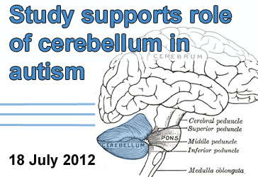
Background: Scientists have long been searching for neural circuits underlying ASD. One suspected player is the cerebellum, a brain region important for balance and movement. Among the various pieces of evidence linking the cerebellum to ASD is the observation that cerebellar pathology is a common feature of individuals with Tuberous Sclerosis Complex (TSC), a genetic disorder in which many individuals also display symptoms of autism. TSC is caused by variants of the genes TSC1 and TSC2, which inhibit signaling of the mTOR pathway.
What’s New: Researchers genetically engineered mice which did not express TSC1 in their cerebellum. These mice displayed ASD-like behaviors, including abnormal social interaction and repetitive behavior/vocalizations. They also showed abnormal function of Purkinje cells, a major cell type of the cerebellum. Treatment of the mice with rapamycin, a drug which inhibits mTOR signaling, prevented the ASD-like behaviors and cerebellar dysfunction.
Why it’s Important: This evidence provides support for the role of the cerebellum in ASD. It also demonstrates roles for TSC1 in cerebellar function.
Help me understand :
| Source(s) : |
| Tweet |
SHANK Research Reveals Important Clues about Autism
By Catherine Croft Swanwick, Ph.D. on June 20, 2012

Overview: Autism has long been thought to result from abnormal development of brain connections called synapses, but new research highlights the SHANK family as key synaptic players in this spectrum of disorders.
Background: SHANK 1, 2, and 3 are “scaffolding proteins,” so called because they literally hold together synaptic components. Many of these synaptic molecules include neurotransmitter receptors that bind to glutamate to produce an excitatory signal in the brain. SHANK2 and SHANK3 identified as ASD risk genes in 2010 and 2007, respectively.
What's New:
1) Mutations of all SHANK family members have now been linked to Autism Spectrum Disorders (ASD). The May 2012 issue of the American Journal of Human Genetics reported genetic evidence gathered from 1,158 Canadian and 456 European individuals with ASD, compared with >15,000 controls. The researchers found two types of SHANK1 mutations associated with ASD in males but not females.
2) Mouse models of ASD were recently created by manipulating the SHANK2 gene. Both models, published in Nature in June 2012, showed behaviors similar to core symptoms of ASD such as abnormal social interaction, reduced communication, and repetitive grooming or jumping. These behaviors also correlated with weaker synaptic function of excitatory neurotransmitter receptors, presumably because their SHANK2 synaptic scaffolds are missing.
Why It’s Important: Together, emerging evidence from SHANK-related research is paving the way for discovery of new targeted ASD drug treatments. For example, to test for potential new ASD treatments, one of these research groups treated their ASD mouse model with drugs that activate excitatory neurotransmitter receptors. They were able to restore normal social interaction by stimulating a type of excitatory neurotransmitter receptor called the NMDA receptor, either directly with D-cycloserine, or indirectly with an activator of another type of excitatory neurotransmitter receptor called the mGluR5 receptor.
Help me understand :
| Source(s) : |
| Tweet |
Strategy to Treat Angelman Syndrome Tested
By Shana R. Spindler, Ph.D. on January 4, 2012
Overview: In a recent issue of Nature, researchers report a therapeutic strategy to treat Angelman syndrome, a developmental disorder related to autism.
Background: Angelman syndrome is caused by a defective version of the UBE3A protein, which helps tag other proteins for elimination from the cell. Individuals get one paternal and one maternal copy of the UBE3A gene, but through a process known as imprinting, only the maternal copy is expressed. Normally, even if the maternal copy of the gene is nonfunctional, the paternal UBE3A remains silent.
What's new: Researchers recently identified a drug, called Topotecan, that allows cells to express the paternal copy of UBE3A by inhibiting part of the cellular machinery that winds and unwinds DNA. After injecting very small amounts of Topotecan into the brains of Angelman syndrome mouse models, the researchers found that neurons from various areas of the brain began making the UBE3A protein from the paternal copy of the gene.
Why it's important: Topotecan is a promising therapeutic strategy for treating Angelman syndrome, suggest the authors. However, the researchers also stress the importance of additional studies to investigate the off-target affects of the drug.
Help me understand :
| Source(s) : |
| Tweet |